Level:
Correct: 0
Incorrect: 0
Marks: 0/0

The heart and blood vessels

Practise 17 exam-style questions on The heart and blood vessels for AQA GCSE Combined Science (Biology Paper 1). Write your answer as you would in the exam and get it marked in seconds against the mark scheme, point by point, with the Study Helper on hand if you get stuck.

  • 17 questions
  • 1–6 marks each
  • Instant marking
  • No sign-up needed
17 questions, marked as you go

Sample questions with model answers

Here are 4 of the questions in this topic, each with a model answer, an explanation of why it earns the marks and the mark scheme an examiner would use.

Question 1

2 marks

The human heart pumps blood around the body in a double circulatory system.

Explain what is meant by a double circulatory system.

Model answer

In a double circulatory system the blood passes through the heart twice on each complete journey around the body. One circuit carries blood from the right ventricle to the lungs and back, and the other carries blood from the left ventricle to the rest of the body and back.

Explanation

The two circuits are joined at the heart. After picking up oxygen in the lungs, blood returns to the heart to be pumped again, at high pressure, to the rest of the body.

Mark scheme
  1. 1 mark

    blood passes through the heart twice for each complete circuit of the body

  2. 1 mark

    one circuit takes blood to the lungs and the other takes blood to the rest of the body

    allow the right side pumps blood to the lungs and the left side pumps blood to the body

Question 2

3 marks

Name the blood vessel that carries out each of these jobs.

Carries deoxygenated blood from the body into the heart.
Carries blood from the heart to the lungs.
Carries oxygenated blood from the lungs back to the heart.

Model answer

From the body into the heart: the vena cava
From the heart to the lungs: the pulmonary artery
From the lungs back to the heart: the pulmonary vein

Explanation

Arteries carry blood away from the heart and veins carry blood towards it. The pulmonary artery is the only artery carrying deoxygenated blood, and the pulmonary vein the only vein carrying oxygenated blood.

Mark scheme
  1. 1 mark

    vena cava

  2. 1 mark

    pulmonary artery

    do not accept pulmonary vein

  3. 1 mark

    pulmonary vein

    do not accept pulmonary artery

Question 3

3 marks

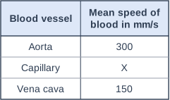
The table shows the mean speed of blood in some blood vessels. Blood takes 1.25 s to flow through a capillary that is 0.5 mm long.

Calculate the speed of blood in the capillary, X.
Calculate how many times faster blood flows in the aorta than in the capillary.

Table with columns "Blood vessel", "Mean speed of blood in mm/s". Blood vessel: Aorta; Mean speed of blood in mm/s: 300 | Blood vessel: Capillary; Mean speed of blood in mm/s: X | Blood vessel: Vena cava; Mean speed of blood in mm/s: 150.

Model answer

Speed in capillary = distance ÷ time = 0.5 ÷ 1.25 = 0.4 mm/s
Aorta ÷ capillary = 300 ÷ 0.4 = 750 times faster

Explanation

Blood flows very slowly in capillaries, which gives more time for substances to diffuse between the blood and the cells. Speed = distance ÷ time.

Mark scheme
  1. 1 mark

    speed = 0.5 ÷ 1.25

    allow distance ÷ time

  2. 1 mark

    = 0.4 (mm/s)

  3. 1 mark

    300 ÷ 0.4 = 750 (times faster)

    allow ecf from an incorrect speed

Question 4

4 marks

A baby is born with a hole in the wall that separates the left and right ventricles.

Suggest and explain how this would affect the baby.

Model answer

Blood would pass through the hole between the ventricles, so oxygenated blood from the left side would mix with deoxygenated blood from the right side. The blood pumped through the aorta to the body would carry less oxygen, so the body cells would get less oxygen for aerobic respiration and less energy would be released. The baby might be tired and breathless and grow slowly, and the heart would have to work harder to supply enough oxygen.

Explanation

In a healthy heart the two sides are completely separate, so blood going to the body is fully oxygenated. Any mixing reduces the oxygen delivered to the tissues.

Mark scheme
  1. 1 mark

    oxygenated and deoxygenated blood mix

    max 4

  2. 1 mark

    so the blood pumped to the body carries less oxygen

  3. 1 mark

    so less oxygen reaches the body cells for (aerobic) respiration

  4. 1 mark

    so less energy is released / the baby would be tired / breathless / grow slowly

    allow skin may look blue

  5. 1 mark

    the heart has to work harder to supply enough oxygen

Ready to try the rest? Every answer you write is marked against the mark scheme, so you can see exactly where the marks are.

Revision notes

The heart and blood vessels

The big idea

The heart is an organ that pumps blood around the body through three types of blood vessel. On each trip round the body the blood also passes through the lungs, where it picks up oxygen and gets rid of carbon dioxide. You need to know the route the blood takes, how the lungs are adapted for gas exchange, and how each vessel's structure suits its job.

The heart and the double circulation

Diagram of a vertical section through the human heart, seen from the front (the heart's right side is on the left of the drawing). The four chambers are shown: the right atrium (upper left of the drawing) above the right ventricle (lower left), and the left atrium (upper right) above the left ventricle (lower right). The wall of the left ventricle is much thicker than the wall of the right ventricle, and the septum separates the two sides. Valves (flaps held by thin tendons) lie between each atrium and its ventricle, and small valves sit at the bases of the pulmonary artery and the aorta. The vena cava enters the top of the right atrium; the pulmonary artery leaves the top of the right ventricle and divides into two branches (to the lungs); two pulmonary veins enter the left atrium from the right; the aorta leaves the top of the left ventricle and arches over the top of the heart. A coronary artery runs over the outer surface of the heart wall over the left ventricle, with small branches. Coloured arrows show the direction of blood flow. Deoxygenated blood (shaded pale blue, blue arrows) flows from the vena cava into the right atrium, down through the valve into the right ventricle, and out through the pulmonary artery to the lungs. Oxygenated blood (shaded pale red, red arrows) flows from the pulmonary veins into the left atrium, down through the valve into the left ventricle, and out through the aorta to the body. The label "vena cava" points to the vena cava; the label "right atrium" points to the right atrium; the label "right ventricle" points to the right ventricle; the label "pulmonary artery" points to the pulmonary artery; the label "aorta" points to the aorta; the label "pulmonary vein" points to the pulmonary vein; the label "left atrium" points to the left atrium; the label "left ventricle" points to the left ventricle; the label "coronary artery" points to the coronary artery.
The heart seen from the front, so its right side is on the left of the drawing. Blue arrows show deoxygenated blood and red arrows oxygenated blood.

Humans have a double circulatory system: blood passes through the heart twice for each complete circuit of the body.

  • The right ventricle pumps blood to the lungs, where gas exchange takes place.
  • The left ventricle pumps blood around the rest of the body.

The route to learn: vena cava \(\rightarrow\) right atrium \(\rightarrow\) right ventricle \(\rightarrow\) pulmonary artery \(\rightarrow\) lungs \(\rightarrow\) pulmonary vein \(\rightarrow\) left atrium \(\rightarrow\) left ventricle \(\rightarrow\) aorta.

The atria receive blood; the ventricles pump it out. Valves stop blood flowing backwards (you do not need their names). The septum keeps oxygenated and deoxygenated blood apart. The left ventricle wall is thicker because it pumps blood at a higher pressure all round the body; both ventricles pump the same volume of blood.

The heart wall is muscle, and it needs its own supply of oxygen and glucose for respiration. This comes through the coronary arteries, because the wall is too thick for enough oxygen to diffuse in from the blood inside the chambers.

The natural resting heart rate is controlled by a group of cells in the right atrium that act as a pacemaker, sending out electrical impulses that make the heart muscle contract. An artificial pacemaker is an electrical device fitted under the skin to correct an irregular heart rate.

The lungs and gas exchange

Air travels down the trachea, into the two bronchi (one to each lung), and on to millions of tiny air sacs called alveoli. Each alveolus is surrounded by a network of capillaries. Oxygen diffuses from the air in the alveoli into the blood, and carbon dioxide diffuses from the blood into the alveoli.

The lungs are adapted for fast diffusion:

  • millions of alveoli give a very large surface area;
  • the alveolus wall and the capillary wall are each one cell thick, so the diffusion distance is short;
  • the blood flow carries oxygen away and breathing brings in fresh air, which keeps a steep concentration gradient.

Arteries, veins and capillaries

Diagram of an artery, a vein and a capillary in cross-section, side by side. The artery (left) has a thick wall (a thin outer layer, a thick middle layer of muscle and elastic fibres with wavy fibres drawn in it, and a thin lining) round a narrow lumen. The vein (middle) has a thin wall (with only a thin muscle layer) round a wide lumen; its outline is slightly flattened. The capillary (right) has a wall only one cell thick round a lumen just wide enough for one red blood cell (drawn inside it); it is drawn much smaller than the others. Below the vein, a short length of vein is drawn cut lengthways (captioned "cut lengthways"), showing a valve: a pair of pocket-shaped flaps. A note says "Not to scale". The label "artery" points to the artery; the label "vein" points to the vein; the label "capillary" points to the capillary; the label "muscle layer" points to the thick layer of muscle and elastic fibres in the artery wall; the label "valve" points to the valve.
An artery, a vein and a capillary in cross-section, with a vein cut lengthways to show a valve.
VesselJobStructure and why
ArteryCarries blood away from the heart at high pressureThick muscular wall to withstand the pressure; elastic fibres stretch and recoil with each heartbeat
VeinCarries blood back to the heart at low pressureThin wall; wide lumen (the space inside) to reduce resistance; valves stop blood flowing backwards
CapillaryExchanges substances with body cellsWall one cell thick for a short diffusion distance; huge networks close to every cell

Arteries and veins are named by direction, not by oxygen: the pulmonary artery carries deoxygenated blood, and the pulmonary vein carries oxygenated blood.

Rates for the heart and blood flow

A student counts 23 pulse beats in 20 seconds. What is her heart rate?

There are three lots of 20 s in a minute: \(23 \times 3 = 69\) beats per minute

In 3 minutes, 2.7 dm³ of blood flows through an organ. Calculate the rate of flow in cm³ per second.

Convert: \(2.7\text{ dm}^3 = 2700\text{ cm}^3\) and \(3\text{ min} = 180\text{ s}\)

\(\text{rate} = \dfrac{\text{volume}}{\text{time}} = \dfrac{2700}{180} = 15\text{ cm}^3\text{ per second}\)

Common mistakes

  • Mixing up the sides. Diagrams show the heart as if you are facing the person, so the right side of the heart is on the left of the drawing.
  • Saying arteries carry oxygenated blood. Arteries carry blood away from the heart; the pulmonary artery carries deoxygenated blood.
  • Saying the left ventricle pumps more blood. It pumps the same volume at a higher pressure.
  • Saying a vein has "bigger" walls or is "bigger". Veins have thin walls and a wide lumen.
  • Subtracting in "how many times thicker" questions. Divide one value by the other.

Exam tip

For "explain how the structure of each vessel relates to its function", link every feature to a reason: "thick walls to withstand high pressure", "valves to prevent backflow", "walls one cell thick for a short diffusion distance". A feature with no reason earns little credit.

Study Helper Hints first, answers after you try

Stuck on this one? Ask for a hint and I’ll point you in the right direction, without giving the answer away. Once you’ve checked your answer, I’ll show you how the marks are awarded.