Level:
Correct: 0
Incorrect: 0
Marks: 0/0

Blood

Practise 11 exam-style questions on Blood for AQA GCSE Combined Science (Biology Paper 1). Write your answer as you would in the exam and get it marked in seconds against the mark scheme, point by point, with the Study Helper on hand if you get stuck.

  • 11 questions
  • 1–6 marks each
  • Instant marking
  • No sign-up needed
11 questions, marked as you go

Sample questions with model answers

Here are 4 of the questions in this topic, each with a model answer, an explanation of why it earns the marks and the mark scheme an examiner would use.

Question 1

2 marks

Describe the function of platelets.

Model answer

Platelets help the blood to clot at the site of a wound. The clot stops bleeding and prevents microorganisms from entering the body.

Explanation

Platelets are small fragments of cells with no nucleus. If someone has too few platelets, they bruise easily and cuts take a long time to stop bleeding.

Mark scheme
  1. 1 mark

    they help the blood to clot

    allow form clots / scabs

  2. 1 mark

    at a wound, to stop bleeding / to stop microorganisms getting in

Question 2

2 marks

A red blood cell has a diameter of 7.5 μm. In a photograph taken through a microscope, the image of the cell has a diameter of 30 mm.

Calculate the magnification of the photograph.

Model answer

30 mm = 30 000 μm
Magnification = size of image ÷ size of real object = 30 000 ÷ 7.5 = ×4000

Explanation

Convert both sizes to the same unit before dividing. Magnification has no unit.

Mark scheme
  1. 1 mark

    30 mm = 30 000 μm and 30 000 ÷ 7.5

    allow 7.5 μm = 0.0075 mm and 30 ÷ 0.0075; an answer of ×4000 scores 2 marks

  2. 1 mark

    ×4000

    allow 4000; allow 4 × 10³

Question 3

3 marks

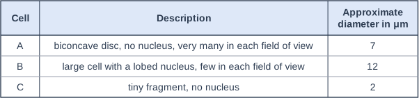
A student looked at a blood smear with a light microscope. The table shows a description of each of three types of cell, A, B and C, that she saw.

Identify A, B and C.

Table with columns "Cell", "Description", "Approximate diameter in μm". Cell: A; Description: biconcave disc, no nucleus, very many in each field of view; Approximate diameter in μm: 7 | Cell: B; Description: large cell with a lobed nucleus, few in each field of view; Approximate diameter in μm: 12 | Cell: C; Description: tiny fragment, no nucleus; Approximate diameter in μm: 2.

Model answer

A is a red blood cell, B is a white blood cell and C is a platelet.

Explanation

Red blood cells are biconcave discs with no nucleus. White blood cells are larger and have a nucleus, which is often lobed. Platelets are tiny fragments of cells with no nucleus, and they are much smaller than red blood cells.

Mark scheme
  1. 1 mark

    A: red blood cell

  2. 1 mark

    B: white blood cell

    allow phagocyte / lymphocyte

  3. 1 mark

    C: platelet

Question 4

4 marks

A person with anaemia has fewer red blood cells than normal. They often feel tired and become breathless easily.

Explain why.

Model answer

With fewer red blood cells there is less haemoglobin, so the blood carries less oxygen to the body's cells. The cells, especially the muscles, cannot carry out as much aerobic respiration, so less energy is released and the person feels tired. The person becomes breathless because they breathe faster and more deeply to try to get more oxygen into the blood.

Explanation

Link the shortage of red blood cells to oxygen transport, then to respiration and energy. Breathlessness is the body's attempt to make up for the lower oxygen supply.

Mark scheme
  1. 1 mark

    less haemoglobin, so the blood carries less oxygen

  2. 1 mark

    less oxygen reaches the body cells / muscles

  3. 1 mark

    so less aerobic respiration happens and less energy is released (so they feel tired)

  4. 1 mark

    they breathe faster / become breathless to try to take in more oxygen

Ready to try the rest? Every answer you write is marked against the mark scheme, so you can see exactly where the marks are.

Revision notes

Blood

The big idea

Blood is a tissue. It is made of a liquid, the plasma, in which red blood cells, white blood cells and platelets are suspended. Each component has its own job: transporting substances, carrying oxygen, defending the body against pathogens or helping the blood to clot. You need to know each job, recognise each part in a diagram or photograph, and explain how the cells are adapted.

The four components

Diagram of a sample of blood seen under a microscope (template blood_sample). A blood smear seen under a microscope: plasma (the pale yellow liquid), 12 red blood cells (biconcave discs with no nucleus; one is seen edge-on, showing the biconcave shape), one white blood cell (larger, with a lobed nucleus) and 4 platelets (small cell fragments). The label "red blood cell" points to a red blood cell (biconcave disc with no nucleus); the label "white blood cell" points to the white blood cell; the label "platelet" points to a platelet (a small cell fragment); the label "plasma" points to the plasma.
Blood seen under a light microscope: red blood cells are by far the most common cells.
ComponentWhat it looks likeFunction
PlasmaPale yellow liquid, mostly waterTransports dissolved substances around the body
Red blood cellsSmall biconcave discs with no nucleus; very many of themCarry oxygen from the lungs to the body cells
White blood cellsLarger, fewer, with a nucleus (often lobed)Defend the body against pathogens
PlateletsTiny fragments of cells with no nucleusHelp the blood to clot at a wound

Substances carried in the plasma include:

  • carbon dioxide from the organs to the lungs;
  • soluble products of digestion (such as glucose and amino acids) from the small intestine to other organs;
  • urea from the liver to the kidneys;
  • hormones, such as insulin, from glands to their target organs.

How the blood cells are adapted

Red blood cells

  • They are biconcave (thinner in the middle), which gives a large surface area for oxygen to diffuse in and out quickly.
  • They have no nucleus, which leaves more room for haemoglobin.
  • Haemoglobin is a red protein that combines with oxygen in the lungs to form oxyhaemoglobin, then releases the oxygen in the body tissues.
  • They are small and flexible, so they can squeeze through narrow capillaries close to body cells.

Because they have no nucleus, mature red blood cells cannot divide; new ones are made in the bone marrow.

White blood cells are part of the immune system. They protect the body in three ways:

  1. Phagocytosis: some can change shape to engulf pathogens and then digest them inside the cell.
  2. Producing antibodies, which target particular pathogens.
  3. Producing antitoxins, which counteract the toxins that some bacteria release.

Platelets help to form a clot that plugs a damaged blood vessel. A clot prevents blood loss and stops pathogens getting into the wound, and it dries to form a scab while the skin underneath heals.

Using blood data

Blood tests compare a person's counts with a healthy range. Link each result to the function it affects:

  • too few red blood cells (or too little haemoglobin, as in anaemia): less oxygen reaches the cells, so less aerobic respiration and less energy released, making the person tired and breathless;
  • too few working white blood cells: more frequent or more serious infections;
  • too few platelets: blood clots slowly, so cuts bleed for longer and bruises form easily.

Finding the real size of a blood cell

In a drawing magnified \(\times 3000\), a platelet is 9 mm wide. Calculate its real width in micrometres.

Rearrange: \(\text{real size} = \dfrac{\text{size of image}}{\text{magnification}} = \dfrac{9}{3000} = 0.003\text{ mm}\)

Convert: \(0.003 \times 1000 = 3\ \mu\text{m}\)

Remember

  • Blood is a tissue: plasma plus red blood cells, white blood cells and platelets.
  • Red blood cell: biconcave, no nucleus, full of haemoglobin, carries oxygen.
  • White blood cells: phagocytosis, antibodies, antitoxins.
  • Platelets: fragments of cells that help the blood clot.

Common mistakes

  • Writing "blood cell" or "red cell" on its own. Name it fully: red blood cell or white blood cell.
  • Saying haemoglobin "absorbs" oxygen. Haemoglobin combines with (binds to) oxygen.
  • Saying white blood cells "are antibodies" or "make antigens". They produce antibodies; antigens are on the pathogen.
  • Saying white blood cells "fight germs". Say how: engulf and digest pathogens, or produce antibodies or antitoxins.
  • Saying plasma carries urine to the kidneys. It carries urea; the kidneys make urine.

Exam tip

"Compare" questions need both cells in every point, joined by "but" or "whereas": "red blood cells have no nucleus, but white blood cells do". Describing each cell separately can lose marks. When asked how you identified a cell in a diagram, give a visible feature, such as "it has a nucleus", not its colour.

Study Helper Hints first, answers after you try

Stuck on this one? Ask for a hint and I’ll point you in the right direction, without giving the answer away. Once you’ve checked your answer, I’ll show you how the marks are awarded.