
Level:
Correct: 0
Incorrect: 0
Marks: 0/0
Exam Practice
Course:
Topics:
AQA A-Level Biology (Paper 1) Smart practice A mixed round aimed at your weak spots
Unit 1
Biological molecules
Unit 2
Cells
Unit 3
Organisms exchange substances with their environment
Unit 4
Genetic information, variation and relationships between organisms
DNA, genes and chromosomes
0%
Genetic diversity can arise as a result of mutation or during meiosis
Gene mutations and chromosome non-disjunction
0%
Meiosis and genetic variation
0%
Genetic diversity and adaptation
0%
Species and taxonomy
0%
Biodiversity within a community
0%
Investigating diversity
0%
Avg
score:
–
Topics
done:
0/35

Blood vessels and tissue fluid

Practise 17 exam-style questions on Blood vessels and tissue fluid for AQA A-Level Biology (Paper 1). Write your answer as you would in the exam and get it marked in seconds against the mark scheme, point by point, with the Study Helper on hand if you get stuck.

  • 17 questions
  • 1–8 marks each
  • Instant marking
  • No sign-up needed
17 questions, marked as you go

Sample questions with model answers

Here are 4 of the questions in this topic, each with a model answer, an explanation of why it earns the marks and the mark scheme an examiner would use.

Question 1

2 marks

In varicose veins, the valves in the veins of the legs do not close properly. The veins become swollen and twisted.

Explain why damaged valves cause the veins of the legs to swell.

Model answer

Valves normally stop blood in the leg veins from flowing backwards. When they do not close properly, blood flows back down the leg under gravity and collects in the veins. The extra blood raises the pressure in the veins and stretches their thin walls, which have little muscle or elastic tissue to resist it, so the veins swell.

Explanation

Blood in leg veins is at low pressure and has to travel upwards, so valves are essential to keep it moving one way. Without working valves, blood pools in the veins, and their thin walls stretch. Answers should link the faulty valves to backflow and then to pooling or raised pressure.

Mark scheme
  1. 1 mark

    Valves normally prevent backflow; when damaged, blood flows backwards / down the leg (because of gravity)

    Max 2.

  2. 1 mark

    (So) blood collects/pools in the veins (of the lower leg)

  3. 1 mark

    (So) the pressure in the veins increases and stretches their thin walls (which have little muscle or elastic tissue)
Question 2

3 marks

The rate of blood flow through a blood vessel is proportional to the fourth power of its radius (radius⁴), if the blood pressure stays the same.

Scientists applied a drug that relaxes smooth muscle to an arteriole in a sample of living tissue. The radius of the arteriole increased from 15 μm to 18 μm.

Calculate how many times greater the rate of blood flow through the arteriole became. Give your answer to 2 significant figures.

Use your answer to explain why changing the diameter of arterioles is an effective way to control the flow of blood to a tissue.

Model answer

Flow increases by (18 ÷ 15)⁴ = 1.2⁴ = 2.0736 = 2.1 times (2 s.f.)

The radius increased by only 20%, but the blood flow roughly doubled. So a small amount of contraction or relaxation of the smooth muscle in an arteriole wall causes a large change in the blood flow to a tissue, which makes arterioles a very effective way of controlling where blood goes.

Explanation

Because flow depends on radius to the fourth power, find the ratio of the radii (18 ÷ 15 = 1.2) and raise it to the fourth power, or work out 18⁴ ÷ 15⁴. Forgetting the power gives 1.2, which is the commonest error. This relationship explains why arterioles, with a thick layer of smooth muscle, are the main vessels that control blood flow into capillary beds.

Mark scheme
  1. 1 mark

    Correct method: (18 ÷ 15)⁴ / 1.2⁴ / 18⁴ ÷ 15⁴; or a correct final answer of 2.1 with no working

    A correct answer of 2.1 (times) with no working scores 2 marks (points 1 and 2). Accept for 1 mark: 2.07 or 2.0736 (not given to 2 significant figures) OR 1.2 (ratio of radii not raised to the fourth power).

  2. 1 mark

    2.1 (times)

  3. 1 mark

    A small change in radius/diameter (a 20% increase) gives a large change in blood flow (about double), so small contractions or relaxations of the smooth muscle control flow effectively

    Accept the converse for constriction. ECF: credit an explanation consistent with the student's own value if it is greater than 1.2.

Question 3

4 marks

Explain how two features of the structure of capillaries make capillary beds efficient exchange surfaces.

Model answer

1. The wall of a capillary is only one cell thick, made of thin, flattened endothelial cells, so the diffusion distance between the blood and the tissue cells is very short.
2. Capillaries are very numerous and highly branched, forming a network through the tissue, so there is a large surface area for exchange and every cell is close to a capillary.

Explanation

Each feature must be followed by how it helps exchange: a short diffusion distance, a large surface area, slow flow giving more time, or gaps that let tissue fluid form. A common error is to describe the capillary wall as 'one cell membrane thick' or to refer to a 'cell wall'. Give exactly two features, each with its explanation.

Mark scheme
  1. 1 mark

    Wall (endothelium) is only one cell thick / made of thin, flattened cells

    Max 4. Mark as pairs: 1 and 2, 3 and 4, 5 and 6, 7 and 8. Each explanation mark needs the feature immediately before it. List rule: if more than two features are given, each wrong or contradictory extra feature cancels one correct pair. Reject 'cell wall' and 'capillary walls are one cell membrane thick'.

  2. 1 mark

    (So) short diffusion distance/pathway (between the blood and the tissue cells)

  3. 1 mark

    Narrow lumen (red blood cells pass through in single file / are squeezed against the wall)

  4. 1 mark

    (So) short diffusion distance (from red blood cells to the tissues) OR blood flows slowly, giving more time for exchange

  5. 1 mark

    Capillaries are numerous / highly branched (form a network)

  6. 1 mark

    (So) large surface area (for exchange) OR every cell is close to a capillary (short diffusion distance)

  7. 1 mark

    Gaps/pores between the endothelial cells

    Accept fenestrations.

  8. 1 mark

    (So) water and small molecules (e.g. glucose) can pass out to form tissue fluid OR white blood cells can leave the blood

Question 4

5 marks

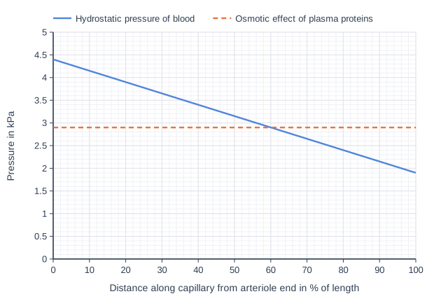
The graph shows two pressures along a capillary in a tissue. The solid line shows the hydrostatic pressure of the blood. The dashed line shows the tendency of water to move into the capillary by osmosis, given as a pressure. This is caused by the lower water potential of the blood due to plasma proteins, and stays about the same along the capillary.

Use the graph to give the distance along the capillary at which the net movement of water changes direction.

Explain the net movement of water at the arteriole end and at the venule end of the capillary.

Use the graph to suggest why more fluid leaves the capillary than returns to it, and state what happens to the excess fluid.

Line graph of pressure in kPa (y-axis, 0 to 5) against distance along capillary from arteriole end in % of length (x-axis, 0 to 100). "Hydrostatic pressure of blood" joins the points (0, 4.4), (100, 1.9). "Osmotic effect of plasma proteins" (dashed) joins the points (0, 2.9), (100, 2.9).

Model answer

The net movement of water changes direction at 60% of the length of the capillary from the arteriole end.
At the arteriole end the hydrostatic pressure (4.4 kPa) is greater than the osmotic effect of the plasma proteins (2.9 kPa), so water and small molecules are forced out into the tissue fluid. At the venule end the hydrostatic pressure (1.9 kPa) is lower than 2.9 kPa, so water moves back into the capillary by osmosis, down the water potential gradient.
Water is forced out along 60% of the capillary, and by a larger pressure difference (up to 1.5 kPa) than it returns (up to 1.0 kPa along 40%), so more fluid leaves than returns. The excess tissue fluid drains into lymph vessels and is returned to the blood.

Explanation

The net direction of water movement depends on which is larger: the hydrostatic pressure pushing water out or the water potential gradient (caused by plasma proteins) drawing it in. Where the lines cross, there is no net movement. Because more fluid leaves than returns along the capillary, the lymph system is needed to return the excess.

Mark scheme
  1. 1 mark

    60 (%) (accept 58 to 62) (of the length from the arteriole end)

  2. 1 mark

    At the arteriole end the hydrostatic pressure (4.4 kPa) is greater than the osmotic effect of the proteins (2.9 kPa), so water (and small molecules) are forced out of the capillary (into the tissue fluid)

    Accept 'hydrostatic pressure is higher than the pressure due to the water potential gradient'.

  3. 1 mark

    At the venule end the hydrostatic pressure (1.9 kPa) is lower than the osmotic effect (2.9 kPa), so water moves into the capillary by osmosis (down a water potential gradient)

    Ignore 'tissue fluid moves in by osmosis': it does not earn this point (it is water that moves by osmosis).

  4. 1 mark

    Water is forced out over a longer distance (the first 60% of the capillary) and/or by a larger pressure difference (up to 1.5 kPa compared with up to 1.0 kPa), so more fluid leaves than returns

    A reason taken from the graph is needed. ECF: accept a distance consistent with the student's own answer for where the lines cross.

  5. 1 mark

    The excess tissue fluid drains into lymph vessels / the lymphatic system (and is returned to the blood)

    Accept lymph capillaries. Ignore 'lymph' or 'lymphatic system' with no idea of the excess fluid draining into it.

Ready to try the rest? Every answer you write is marked against the mark scheme, so you can see exactly where the marks are.

Study Helper Hints first, answers after you try

Stuck on this one? Ask for a hint and I’ll point you in the right direction, without giving the answer away. Once you’ve checked your answer, I’ll show you how the marks are awarded.