
Level:
Correct: 0
Incorrect: 0
Marks: 0/0
Exam Practice
Course:
Topics:
AQA A-Level Biology (Paper 1) Smart practice A mixed round aimed at your weak spots
Unit 1
Biological molecules
Unit 2
Cells
Unit 3
Organisms exchange substances with their environment
Unit 4
Genetic information, variation and relationships between organisms
DNA, genes and chromosomes
0%
Genetic diversity can arise as a result of mutation or during meiosis
Gene mutations and chromosome non-disjunction
0%
Meiosis and genetic variation
0%
Genetic diversity and adaptation
0%
Species and taxonomy
0%
Biodiversity within a community
0%
Investigating diversity
0%
Avg
score:
–
Topics
done:
0/35

Antigens, phagocytosis and T lymphocytes

Practise 18 exam-style questions on Antigens, phagocytosis and T lymphocytes for AQA A-Level Biology (Paper 1). Write your answer as you would in the exam and get it marked in seconds against the mark scheme, point by point, with the Study Helper on hand if you get stuck.

  • 18 questions
  • 1–7 marks each
  • Instant marking
  • No sign-up needed
18 questions, marked as you go

Sample questions with model answers

Here are 4 of the questions in this topic, each with a model answer, an explanation of why it earns the marks and the mark scheme an examiner would use.

Question 1

2 marks

Cytotoxic T cells (TC cells) can destroy cancer cells, which have abnormal proteins on their cell-surface membrane.

Some cancer cells make fewer of the cell-surface proteins that display antigens to T cells. Suggest how this helps these cancer cells to avoid being destroyed.

Model answer

The cancer cell displays fewer of its abnormal antigens on its surface, so it is less likely to be recognised as abnormal. Fewer TC cells with complementary receptors can bind to it, so the cancer cell is not destroyed.

Explanation

TC cells identify abnormal body cells by binding, with their complementary receptors, to the abnormal antigens displayed on the cell surface. If fewer antigens are displayed, fewer TC cells bind and the cancer cell escapes destruction. In an unfamiliar context like this, apply the idea that recognition depends on antigens being present on the cell surface.

Mark scheme
  1. 1 mark

    Fewer (abnormal/non-self) antigens are displayed/presented on the surface of the cancer cell / the cell is not recognised as abnormal

    Accept 'the cancer cell does not present its abnormal antigens'.

  2. 1 mark

    (So) fewer/no TC cells (with complementary receptors) bind to the cancer cell, so it is not killed

    Accept 'TC cells cannot recognise the cancer cell' or 'no perforin is released into it'. Accept as an alternative route to this mark: fewer helper T cells are activated, so there is less clonal expansion of TC cells.
Question 2

3 marks

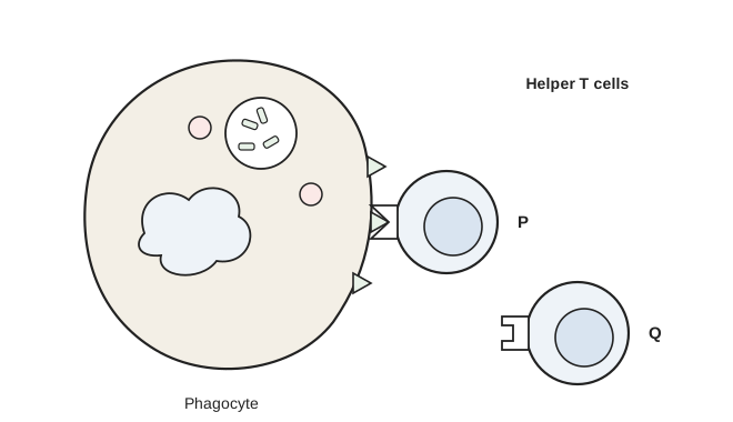
The diagram shows a phagocyte that has engulfed and digested a pathogen, and two helper T cells, P and Q.

Name the role that the phagocyte is now carrying out. Explain which helper T cell, P or Q, will be activated, and describe what this T cell will do next.

A large cell labelled Phagocyte contains a lobed nucleus, two small round bodies and a vacuole holding small fragments. Three small triangular shapes stick out from its surface on the right. To the right, under the heading Helper T cells, are two smaller round cells labelled P and Q, each with a receptor on its left side. Cell P sits against one of the triangles: its receptor has a triangular notch into which the triangle fits. Cell Q is further away: its receptor has a rectangular notch.

Model answer

The phagocyte is acting as an antigen-presenting cell: it displays antigens from the pathogen on its cell-surface membrane. Helper T cell P will be activated, because its receptor has a shape complementary to the antigen, so it can bind to it; Q's receptor is not complementary. P then divides rapidly by mitosis to form a clone of identical T cells, which stimulate cytotoxic T cells, B cells and phagocytes.

Explanation

After digesting a pathogen, a phagocyte places the pathogen's antigens on its surface and becomes an antigen-presenting cell. Each helper T cell has receptors of one specific shape, so only the few T cells whose receptors are complementary to that antigen bind and are activated (clonal selection). The activated cell then divides by mitosis (clonal expansion). Say 'complementary', not 'the same shape'.

Mark scheme
  1. 1 mark

    Antigen presentation / it is an antigen-presenting cell (APC)

    Accept 'presenting antigens (on its surface)'. Ignore 'phagocytosis'.

  2. 1 mark

    P, because its receptor is complementary to (the shape of) the antigen

    Both P and the reason are needed. Reject 'P because its receptor is the same shape as the antigen'. Reject reference to an active site.

  3. 1 mark

    (P) divides by mitosis (to form a clone) / clonal expansion

    Accept 'P is cloned'. Accept as an alternative for this mark: P stimulates cytotoxic T cells, B cells or phagocytes.

Question 3

4 marks

Trypanosomes are single-celled parasites that live in human blood. Each trypanosome is covered by a coat made of one type of glycoprotein. A trypanosome has genes for many different coat glycoproteins but uses only one at a time. A few trypanosomes in a population switch to making a different coat glycoprotein.

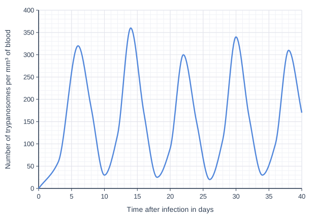
The graph shows the number of trypanosomes in the blood of an infected person over 40 days.

Use the information and your knowledge of antigens and the immune response to explain the pattern shown in the graph.

Suggest why it has not been possible to make an effective vaccine against trypanosomes.

Line graph of number of trypanosomes per mm³ of blood (y-axis, 0 to 400) against time after infection in days (x-axis, 0 to 40). The line is a smooth curve through (0, 0), (3, 60), (6, 320), (8, 180), (10, 30), (12, 120), (14, 360), (16, 170), (18, 25), (20, 90), (22, 300), (24, 150), (26, 20), (28, 110), (30, 340), (32, 160), (34, 30), (36, 100), (38, 310), (40, 170).

Model answer

The number of trypanosomes rises to a peak (about 320 per mm³ at day 6) because they divide in the blood before the immune system has made enough antibodies. B cells with receptors complementary to the coat glycoprotein are then cloned and plasma cells release antibodies, so trypanosomes with that coat are destroyed and the number falls (to about 30 per mm³ at day 10). A few trypanosomes have switched to a different coat glycoprotein, which is a different antigen; the antibodies and memory cells are not complementary to it, so these trypanosomes survive and multiply, producing the next peak. Each new coat needs a new primary response, which takes several days, so the peaks repeat about every 8 days.

A vaccine could contain only one or a few of the coat glycoproteins, so memory cells would not recognise trypanosomes that had switched to a different coat.

Explanation

This is antigen variability in an unfamiliar organism. Each fall in numbers is the immune response removing trypanosomes with the current coat; each new rise is a sub-population with a new coat antigen, which the existing antibodies and memory cells do not fit, so a new primary response is needed every time. The same reasoning explains why vaccines are hard to make against pathogens whose antigens keep changing.

Mark scheme
  1. 1 mark

    Number increases as trypanosomes divide/reproduce (before enough antibodies/an immune response has developed)

    Max 4. Max 3 from points 1 to 5 (point 6 is needed for full marks).

  2. 1 mark

    (After each peak) number falls as antibodies/the immune response destroys trypanosomes with that coat glycoprotein/antigen

    Accept 'plasma cells produce antibodies complementary to the coat'.

  3. 1 mark

    A few trypanosomes have switched to a different glycoprotein/antigen, so the antibodies/memory cells are not complementary to them and they survive

    Accept 'antigen variability' with an explanation.

  4. 1 mark

    These trypanosomes multiply, producing the next peak, and a new primary response is needed for each new antigen (which takes several days)

  5. 1 mark

    Peaks occur about every 8 days (at about days 6, 14, 22, 30 and 38)

    Accept any correct reference to the timing or values of the peaks or troughs.

  6. 1 mark

    (Vaccine) a vaccine would contain only one/a few of the coat glycoproteins, so trypanosomes with other coats would not be recognised by memory cells/antibodies

    Accept 'there are too many different coat antigens to include in a vaccine'.

Question 4

6 marks

A virus infects cells lining the airways. Describe the cellular response that leads to the destruction of the infected cells.

Start your answer from the point when a phagocyte has engulfed and digested some of the virus particles. Do not describe the response of B cells.

Model answer

The phagocyte presents antigens from the virus on its cell-surface membrane, so it is an antigen-presenting cell. A helper T cell with a receptor complementary to one of these antigens binds to it and is activated. The TH cell divides rapidly by mitosis to form a clone of identical TH cells. These TH cells stimulate phagocytes and activate cytotoxic T cells (TC cells) with receptors complementary to the same antigen, which also divide by mitosis to form a clone. Infected airway cells display viral antigens on their cell-surface membranes. TC cells bind to these antigens and kill the infected cells, for example by releasing perforin, which makes holes in the cell-surface membrane. Some of the T cells remain as memory cells.

Explanation

The cellular response is the response of T lymphocytes to antigens on cells: antigen presentation, activation of the specific TH cells, clonal expansion by mitosis, then TH cells stimulating TC cells, which kill infected body cells. A common error is to say that TC cells produce antibodies or engulf cells: they kill infected cells, while antibodies come from plasma cells. Perforin is credited but is not required by the specification.

Mark scheme
  1. 1 mark

    Phagocyte presents viral antigens on its cell-surface membrane / acts as an antigen-presenting cell

    Max 6.

  2. 1 mark

    Helper T cell with a (specific) receptor complementary to the antigen binds to it

    Reject 'same shape' for complementary.

  3. 1 mark

    TH cell (is activated and) divides by mitosis to form a clone / clonal expansion

    Accept 'is cloned'.

  4. 1 mark

    (Activated) TH cells stimulate cytotoxic T cells/TC cells

    Accept 'release chemicals/cytokines that activate TC cells'.

  5. 1 mark

    TC cells (with complementary receptors) divide by mitosis / are cloned

  6. 1 mark

    Infected cells display viral antigens on their cell-surface membrane

  7. 1 mark

    TC cells bind to (the viral antigens on) infected cells

  8. 1 mark

    TC cells kill the infected cells (e.g. by releasing perforin, which makes holes in the cell-surface membrane)

    Perforin is credited but not required. Reject 'TC cells engulf the infected cells' or 'TC cells release antibodies'.

  9. 1 mark

    (Activated) TH cells stimulate phagocytes (to engulf more pathogens/infected cells)

  10. 1 mark

    Some T cells develop into memory cells

    Ignore any description of B cells or antibodies (the question excludes them).

Ready to try the rest? Every answer you write is marked against the mark scheme, so you can see exactly where the marks are.

Study Helper Hints first, answers after you try

Stuck on this one? Ask for a hint and I’ll point you in the right direction, without giving the answer away. Once you’ve checked your answer, I’ll show you how the marks are awarded.