Level:
Correct: 0
Incorrect: 0
Marks: 0/0
Exam Practice
Course:
Topics:
AQA A-Level Biology (Paper 2) Smart practice A mixed round aimed at your weak spots
Unit 5
Energy transfers in and between organisms
Unit 6
Organisms respond to changes in their internal and external environments
Stimuli, both internal and external, are detected and lead to a response
Survival and response
0%
Receptors
0%
Control of heart rate
0%
Skeletal muscles are stimulated to contract by nerves and act as effectors
Skeletal muscles and muscle contraction
0%
Unit 7
Genetics, populations, evolution and ecosystems
Unit 8
The control of gene expression
Alteration of the sequence of bases in DNA can alter the structure of proteins
Gene mutations and their effects on proteins
0%
Using genome projects
0%
Differences in DNA between individuals of the same species can be exploited
DNA probes, screening and genetic counselling
0%
Genetic fingerprinting
0%
Unit 9
Comprehension
Avg
score:
–
Topics
done:
0/34

Receptors

Practise 18 exam-style questions on Receptors for AQA A-Level Biology (Paper 2). Write your answer as you would in the exam and get it marked in seconds against the mark scheme, point by point, with the Study Helper on hand if you get stuck.

  • 18 questions
  • 1–7 marks each
  • Instant marking
  • No sign-up needed
18 questions, marked as you go

Sample questions with model answers

Here are 4 of the questions in this topic, each with a model answer, an explanation of why it earns the marks and the mark scheme an examiner would use.

Question 1

3 marks

The figure shows a Pacinian corpuscle cut lengthways.

Name the type of ion channel in the membrane at X that is responsible for producing a generator potential. Explain why a Pacinian corpuscle produces a generator potential when it is pressed, but not when the skin above it is warmed.
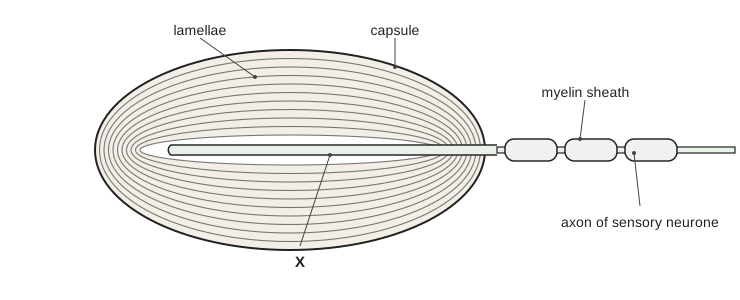
An oval structure cut lengthways, made of many thin layers arranged like the layers of an onion around a narrow central space. A label 'capsule' points to the outer edge and a label 'lamellae' points to the layers. A thin rod-shaped structure runs along the centre of the oval and leaves at its right-hand end, where it continues as a thin fibre covered by a series of rounded segments labelled 'myelin sheath'; the fibre is labelled 'axon of sensory neurone'. Label X points to the surface of the thin rod-shaped structure in the centre of the oval.

Model answer

X contains stretch-mediated sodium ion channels. When the corpuscle is pressed, the lamellae are deformed and the membrane of the neurone ending is stretched. This opens the stretch-mediated sodium ion channels, and sodium ions diffuse in, producing a generator potential. Warming the skin does not deform or stretch the membrane, so the channels stay closed and no sodium ions enter; the corpuscle only responds to its specific stimulus, mechanical pressure.

Explanation

The Pacinian corpuscle is AQA's example of receptor specificity: it responds only to mechanical pressure because its channels open only when the membrane is deformed. Sodium ions then diffuse in, depolarising the membrane to form a generator potential. A common error is to name voltage-gated channels, which are found in the axon of the sensory neurone, not in the receptor membrane.

Mark scheme
  1. 1 mark

    Stretch-mediated sodium (ion) channels

    Both the idea of stretch (or deformation / pressure) and sodium are needed. Reject 'voltage-gated sodium channels' and potassium channels. Accept Na⁺ for sodium ions throughout.

  2. 1 mark

    Pressure deforms the lamellae / stretches the membrane (of the neurone ending), so the channels open AND sodium ions diffuse in

    Accept 'channels widen'. Accept 'sodium ions enter, causing depolarisation'. Reject 'sodium ions are actively transported in'.

  3. 1 mark

    Warming does not deform / stretch the membrane, so the (stretch-mediated) channels stay closed / no sodium ions enter

    Accept 'there are no channels that open in response to temperature'. 'Receptors respond only to specific stimuli' on its own is insufficient: the answer must say why heat has no effect.
Question 2

3 marks

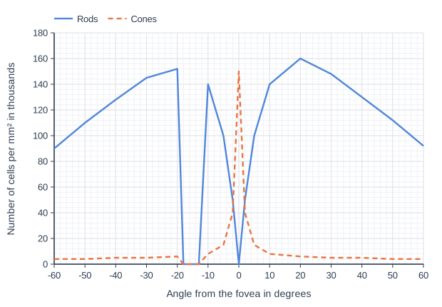
The graph shows the numbers of rod cells and cone cells across the retina of a human eye, measured along a line through the fovea (0°). Negative angles are on the side of the retina nearest the nose.

Calculate the ratio of rod cells to cone cells at 20° from the fovea, on the side away from the nose. Give your answer in the form x : 1, with x to 2 significant figures.

No light can be detected when it falls on the retina between −18° and −13°. Use the graph to explain why.

Line graph of number of cells per mm² in thousands (y-axis, 0 to 180) against angle from the fovea in degrees (x-axis, -60 to 60). "Rods" joins the points (-60, 90), (-50, 110), (-40, 128), (-30, 145), (-20, 152), (-18, 0), (-13, 0), (-10, 140), (-5, 100), (-2, 50), (0, 0), (2, 50), (5, 100), (10, 140), (20, 160), (30, 148), (40, 130), (50, 112), (60, 92). "Cones" (dashed) joins the points (-60, 4), (-50, 4), (-40, 5), (-30, 5), (-20, 6), (-18, 0), (-13, 0), (-10, 8), (-5, 15), (-2, 40), (0, 150), (2, 40), (5, 15), (10, 8), (20, 6), (30, 5), (40, 5), (50, 4), (60, 4).

Model answer

At +20°: rods = 160 thousand per mm², cones = 6 thousand per mm²
Ratio = 160 ÷ 6 = 26.7, so 27 : 1

Between −18° and −13° the graph shows no rods and no cones. This is the blind spot, where the neurones of the optic nerve leave the eye, so light falling there cannot produce generator potentials or impulses.

Explanation

A ratio in the form x : 1 is found by dividing the first number by the second. The blind spot is the region where the optic nerve leaves the retina; it has no photoreceptors, so nothing falling on it is detected. Read both curves carefully and check which side of the fovea the question refers to.

Mark scheme
  1. 1 mark

    27 : 1 (accept 23 : 1 to 32 : 1); a correct answer with no working scores this mark

    Point 1 is the calculation: a correct answer of 27 : 1 (160 ÷ 6 = 26.7) with no working scores 1 mark. Accept answers from 23 : 1 to 32 : 1 (cones read as 5 to 7 thousand per mm², rods as 158 to 162 thousand per mm²). Do not accept 26.7 : 1 (not given to 2 significant figures). Reject 1 : 27 and 0.0375 : 1.

  2. 1 mark

    There are no rods or cones (no receptors) between −18° and −13°

    Accept 'number of rods and cones is 0'.

  3. 1 mark

    So no generator potentials / impulses can be produced there OR this is where the (neurones of the) optic nerve leave the eye / the blind spot

    Accept 'light is not absorbed by any pigment there'.

Question 3

4 marks

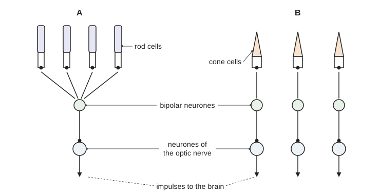
The figure shows how rod cells and cone cells are connected to neurones in two regions of the retina, A and B.

In region A, dim light falls on only one of the four rod cells, and no impulses pass along the neurone of the optic nerve. When the same dim light falls on all four rod cells, impulses pass along this neurone. Explain why.

Two points of light, very close together, fall on two neighbouring cone cells in region B. Explain why the brain can tell that there are two separate points of light.

A diagram of two regions of the retina. In region A, four rod cells in a row at the top each connect to one bipolar neurone below them, and this bipolar neurone connects to one neurone of the optic nerve, from which an arrow labelled impulses to the brain points down. In region B, three cone cells at the top each connect to their own bipolar neurone, and each bipolar neurone connects to its own neurone of the optic nerve, each with an arrow pointing down. The rod cells, cone cells, bipolar neurones and neurones of the optic nerve are labelled.

Model answer

In region A, one rod stimulated by dim light releases too little neurotransmitter onto the bipolar neurone, so its membrane does not reach threshold and no impulse is produced. When all four rods are stimulated, the neurotransmitter they release onto the same bipolar neurone adds together (spatial summation), the threshold is reached and impulses pass along the neurone of the optic nerve.

In region B, each cone is connected to its own bipolar neurone and its own neurone of the optic nerve, so the two neighbouring cones send separate sets of impulses to the brain, which can therefore distinguish two points (high visual acuity).

Explanation

Retinal convergence explains the difference between rods and cones: several rods sharing one bipolar neurone allows spatial summation, so rods give high sensitivity in dim light but low acuity, because the brain cannot tell which rod was stimulated. Cones each have their own connection, so they give high visual acuity. Use the words 'impulses' or 'action potentials', not 'signals' or 'messages'.

Mark scheme
  1. 1 mark

    One rod releases too little neurotransmitter / causes too little depolarisation (of the bipolar neurone), so the threshold is not reached

    Accept 'generator potential in one rod is not enough to reach threshold'. Accept named neurotransmitter.

  2. 1 mark

    With four rods, the neurotransmitter / depolarisations add together / spatial summation, so the threshold is reached (in the bipolar neurone)

    'Summation' alone is insufficient: the idea of several rods adding together, or 'spatial', is needed.

  3. 1 mark

    Each cone is connected to its own bipolar neurone (and its own neurone in the optic nerve)

    Accept 'no retinal convergence'. Accept 'one cone to one neurone'.

  4. 1 mark

    So the two cones send separate (sets of) impulses to the brain

    Reject 'signals' or 'messages' for impulses. Accept action potentials for impulses.

Question 4

5 marks

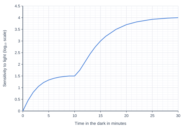
A person spent 30 minutes in bright sunlight and then sat in a completely dark room. Their sensitivity to light was measured at intervals by finding the dimmest flash of light they could see. The graph shows the results. Sensitivity is plotted on a log₁₀ scale: an increase of 1 on the scale means that the eye is 10 times more sensitive.

Bright light breaks down the pigments in rods and in cones. In the dark, the pigments are made again; the pigment in cones is made again faster than the pigment in rods.

Calculate how many times more sensitive to light the eye was after 30 minutes in the dark than after 10 minutes.

Use the information and your knowledge of rods and cones to explain the shape of the curve.

Line graph of sensitivity to light (log₁₀ scale) (y-axis, 0 to 4.5) against time in the dark in minutes (x-axis, 0 to 30). The line joins the points (0, 0), (1, 0.45), (2, 0.8), (3, 1.05), (4, 1.22), (5, 1.33), (6, 1.4), (7, 1.45), (8, 1.48), (9, 1.5), (10, 1.5), (11, 1.75), (12, 2.1), (13, 2.45), (14, 2.75), (15, 3), (16, 3.2), (18, 3.5), (20, 3.7), (22, 3.82), (25, 3.93), (28, 3.98), (30, 4).

Model answer

At 10 minutes the sensitivity is 1.5 and at 30 minutes it is 4.0 on the log₁₀ scale.
Difference = 4.0 − 1.5 = 2.5
Times more sensitive = 10 to the power 2.5 = 316

In the first few minutes sensitivity rises quickly because the cone pigment is made again faster than rhodopsin. By about 7 to 10 minutes it levels off, because cones are not very sensitive: their pigment needs high light intensity and each cone has its own bipolar neurone, so there is no summation. After about 10 minutes enough rhodopsin has been made again for the rods to work, and sensitivity rises again, more slowly, to a much higher level. Rods are far more sensitive because rhodopsin is broken down by low light intensity and several rods share one bipolar neurone, allowing spatial summation.

Explanation

On a log₁₀ scale, subtract the readings and then raise 10 to the power of the difference; dividing the log values (4.0 ÷ 1.5) is a common error. The curve has two parts because cones recover their pigment quickly but have a limited sensitivity, whereas rods recover slowly but end up far more sensitive. This is why it takes about half an hour for your eyes to adjust fully to the dark.

Mark scheme
  1. 1 mark

    Correct method, e.g. 10 to the power (4.0 − 1.5) / 10 to the power 2.5 / the antilog of 2.5; or a correct final answer of 316 with no working

    Max 5. Points 1 and 2 are the calculation: a correct answer of 316 (accept 300 to 320, or 3.2 × 10²) with no working scores 2 marks. Accept for 1 mark: evidence of 2.5 (the difference in log values) not converted. Do not award this mark for 4.0 ÷ 1.5 (2.7). Points 3 to 6: max 3.

  2. 1 mark

    316 (accept 300 to 320)

  3. 1 mark

    0 to 10 minutes: sensitivity increases (quickly) as the pigment in cones (iodopsin) is made again

    Accept 'cones recover first'.

  4. 1 mark

    Sensitivity levels off (at about 6 to 10 minutes) because cones have low sensitivity / cones need high light intensity / each cone has its own neurone (no summation)

    Accept 'the maximum sensitivity of cones is reached'.

  5. 1 mark

    After 10 minutes: sensitivity increases further (more slowly) as rhodopsin in rods is made again / rods start to work

  6. 1 mark

    Rods are much more sensitive because rhodopsin is broken down by low light intensity OR several rods share one bipolar neurone (spatial summation)

Ready to try the rest? Every answer you write is marked against the mark scheme, so you can see exactly where the marks are.

Study Helper Hints first, answers after you try

Stuck on this one? Ask for a hint and I’ll point you in the right direction, without giving the answer away. Once you’ve checked your answer, I’ll show you how the marks are awarded.