Level:
Correct: 0
Incorrect: 0
Marks: 0/0
Exam Practice
Course:
Topics:
AQA A-Level Biology (Paper 2) Smart practice A mixed round aimed at your weak spots
Unit 5
Energy transfers in and between organisms
Unit 6
Organisms respond to changes in their internal and external environments
Stimuli, both internal and external, are detected and lead to a response
Survival and response
0%
Receptors
0%
Control of heart rate
0%
Skeletal muscles are stimulated to contract by nerves and act as effectors
Skeletal muscles and muscle contraction
0%
Unit 7
Genetics, populations, evolution and ecosystems
Unit 8
The control of gene expression
Alteration of the sequence of bases in DNA can alter the structure of proteins
Gene mutations and their effects on proteins
0%
Using genome projects
0%
Differences in DNA between individuals of the same species can be exploited
DNA probes, screening and genetic counselling
0%
Genetic fingerprinting
0%
Unit 9
Comprehension
Avg
score:
–
Topics
done:
0/34

DNA probes, screening and genetic counselling

Practise 18 exam-style questions on DNA probes, screening and genetic counselling for AQA A-Level Biology (Paper 2). Write your answer as you would in the exam and get it marked in seconds against the mark scheme, point by point, with the Study Helper on hand if you get stuck.

  • 18 questions
  • 1–7 marks each
  • Instant marking
  • No sign-up needed
18 questions, marked as you go

Sample questions with model answers

Here are 4 of the questions in this topic, each with a model answer, an explanation of why it earns the marks and the mark scheme an examiner would use.

Question 1

2 marks

People who are found to carry an allele for a heritable condition are often referred to a genetic counsellor.

Describe the role of a genetic counsellor.

Model answer

A genetic counsellor explains the results of screening and how the condition is inherited, including the probability that the person or their children will have or pass on the condition. They explain the options available, such as treatment, further tests or embryo screening, and support the person in making their own informed decision.

Explanation

Genetic counselling uses the information from screening: the counsellor explains what the results mean, the risks to the person and their family, and the choices available. Counsellors do not tell people what to decide; they help them make an informed choice.

Mark scheme
  1. 1 mark

    Explains the results of the screening / what the condition is and how it is inherited

    Max 2. Points 1 to 4: 1 mark each.

  2. 1 mark

    Explains the probability / risk of the person (or their children) having or passing on the condition

  3. 1 mark

    Explains the options available, e.g. treatment, further tests, prenatal testing, embryo screening

  4. 1 mark

    Gives support / helps the person make their own informed decision (without telling them what to do)

    Accept 'discusses the implications for other members of the family'.
Question 2

3 marks

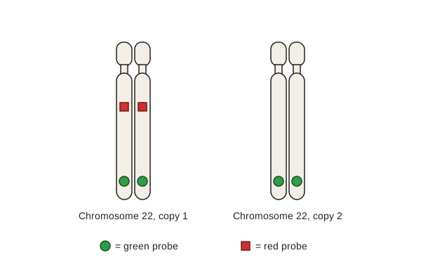
A rare condition is caused by the deletion of a small region from one copy of chromosome 22. Doctors used two fluorescent probes on chromosomes from a child's cells. The green probe binds to a base sequence near the end of chromosome 22. The red probe binds to a base sequence inside the region that is deleted in this condition. The figure shows the results for the two copies of chromosome 22 from one cell.

Explain what the results show about the child's chromosomes 22.

The child's parents were tested in the same way, and neither of them has the deletion. Suggest what this shows.

Two copies of chromosome 22, each drawn as two chromatids joined at a centromere near the top, labelled 'Chromosome 22, copy 1' and 'Chromosome 22, copy 2'. A key shows a green circle for the green probe and a red square for the red probe. Copy 1 has a green circle near the end of the long arm on each chromatid and a red square part of the way along the long arm on each chromatid. Copy 2 has a green circle near the end of the long arm on each chromatid and no red squares.

Model answer

Both copies of chromosome 22 have green signals, so both are present and the probes could bind. The red probe has bound to copy 1 but not to copy 2, so copy 2 is missing the region that contains the red probe's target sequence. The child has the deletion on one copy of chromosome 22.

The deletion must be a new mutation that happened when one parent's gametes were produced (during meiosis), so it was not inherited and the chance of it happening again in another child is low.

Explanation

Fluorescent probes can be used to locate specific base sequences on chromosomes: a missing signal shows that the target sequence is absent. A second probe that binds elsewhere on the same chromosome identifies the chromosome and shows that the test worked. Testing parents shows whether a change was inherited, which is important for genetic counselling.

Mark scheme
  1. 1 mark

    The red probe binds to copy 1 but not to copy 2, so copy 2 does not contain the base sequence / region that the red probe binds to

    Max 3. Points 1 to 3: 1 mark each, max 2 from these points. Point 4: 1 mark. Accept 'copy 2 has no red signal so the region is missing'.

  2. 1 mark

    So the child has the deletion on one copy of chromosome 22 (and so has the condition)

    Accept 'the child is heterozygous for the deletion'.

  3. 1 mark

    The green probe binds to both copies, showing that both copies of chromosome 22 are present / that the probes could bind (so the missing red signal is not due to a failed test)

    Accept 'green shows which chromosomes are chromosome 22'.

  4. 1 mark

    (Parents) the deletion is a new mutation, which happened in a gamete / during meiosis in one parent, so the risk for the couple's future children is low

    Accept 'the deletion was not inherited from either parent'.

Question 3

4 marks

In some breast tumours, the cells contain many copies of gene G, which codes for a receptor that stimulates cell division. Drug H is effective only against tumours in which gene G has been copied many times.

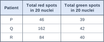
Doctors tested tumour cells from three patients using two fluorescent probes: a red probe that binds to gene G, and a green probe that binds to the centromere of chromosome 17, the chromosome that carries gene G. They counted the red spots and the green spots in 20 nuclei from each tumour. A tumour is treated with drug H if the ratio of red spots to green spots is 2.0 or more. The table shows their results.

Calculate the ratio of red spots to green spots for each patient, and identify which patients should be treated with drug H.

Explain why the doctors used the green probe as well as the red probe.

Suggest why the doctors counted spots in 20 nuclei, rather than in one nucleus.

Table with columns "Patient", "Total red spots in 20 nuclei", "Total green spots in 20 nuclei". Patient: P; Total red spots in 20 nuclei: 46; Total green spots in 20 nuclei: 39 | Patient: Q; Total red spots in 20 nuclei: 162; Total green spots in 20 nuclei: 42 | Patient: R; Total red spots in 20 nuclei: 84; Total green spots in 20 nuclei: 40.

Model answer

P: 46 ÷ 39 = 1.18; Q: 162 ÷ 42 = 3.86; R: 84 ÷ 40 = 2.10
Patients Q and R have ratios of 2.0 or more, so they should be treated with drug H.

Tumour cells often have extra copies of whole chromosomes. The green probe counts the copies of chromosome 17, so the ratio shows whether gene G has been copied more times than the chromosome that carries it.

The number of spots varies from cell to cell, so a mean from 20 nuclei is more representative of the tumour than one nucleus.

Explanation

Fluorescent probes that bind to specific base sequences can be used to count genes in cells, a form of screening that guides personalised medicine. Dividing the gene count by the chromosome count corrects for tumour cells that have extra chromosomes. Results from many cells are needed because individual cells vary.

Mark scheme
  1. 1 mark

    Correct ratios for all three patients: P 1.2 (1.18), Q 3.9 (3.86), R 2.1 (naming Q and R with no working scores point 2 only)

    Max 4. A correct answer naming Q and R with the three correct ratios scores 2 marks (points 1 and 2). Naming Q and R with no working (no ratios) scores 1 mark (point 2 only). Accept ratios to 2 or more significant figures, e.g. 1.18, 3.86, 2.10, and ratios written as 1.2 : 1, 3.9 : 1 and 2.1 : 1. Do not credit unsimplified ratios such as 46 : 39 for point 1.

  2. 1 mark

    Patients Q and R (should be treated with drug H)

    ECF: credit the patients whose ratios, as calculated by the student, are 2.0 or more.

  3. 1 mark

    Tumour cells may have extra copies of chromosome 17, so the green probe shows how many copies there are; the ratio then shows whether gene G itself has been copied more than the chromosome

    Accept 'the green probe acts as a control / reference for the number of chromosomes 17'. Insufficient: 'as a control' unqualified.

  4. 1 mark

    The number of spots varies between cells / nuclei, so a mean of many nuclei is more representative / reliable

    Accept 'one nucleus might not be typical of the tumour' or 'reduces the effect of anomalous nuclei'.

Question 4

6 marks

Describe how a labelled DNA probe could be used to find out whether a person has a particular allele of a gene.

Do not include gel electrophoresis in your answer.

Model answer

The base sequence of the allele must be known. A probe is made: a short, single-stranded length of DNA with a base sequence complementary to part of the allele, labelled with a fluorescent dye or a radioactive isotope. DNA is extracted from the person's cells and amplified by PCR. The DNA is heated to break the hydrogen bonds between the strands, making it single-stranded. The probe is added; if the allele is present, the probe binds (hybridises) to the complementary base sequence by hydrogen bonds between complementary bases. Unbound probe is washed away. The label is then detected - fluorescence under UV light, or radioactivity using X-ray film. A signal shows that the person has the allele.

Explanation

The essential ideas are a single-stranded, labelled probe complementary to the allele; single-stranded target DNA; hybridisation by complementary base pairing; washing off unbound probe; and detecting the label. A common error is to confuse probes with primers or with marker genes. Saying that the probe 'attaches' is not enough: say that it binds to a complementary base sequence.

Mark scheme
  1. 1 mark

    The base sequence of the allele (or part of it) must be known, so a probe with a complementary base sequence can be made

    Max 6. Accept 'probe is complementary to (part of) the allele / gene / DNA'.

  2. 1 mark

    The probe is a short, single-stranded length of DNA

  3. 1 mark

    The probe is labelled with a fluorescent dye / radioactive isotope

    Accept 'fluorescent / radioactive marker'.

  4. 1 mark

    DNA is extracted from the person's cells and copied / amplified by PCR

    Accept 'many copies made using PCR'.

  5. 1 mark

    The DNA is made single-stranded by heating / hydrogen bonds between the strands are broken

    Accept 'denatured' if linked to separating the strands.

  6. 1 mark

    The probe is added and binds / hybridises to the complementary base sequence (if the allele is present), by hydrogen bonds between complementary bases

    Insufficient: 'the probe attaches to the DNA' with no idea of complementary base pairing or hybridisation.

  7. 1 mark

    Unbound probe is washed away

  8. 1 mark

    The label is detected, e.g. fluorescence seen under UV light OR radioactivity detected with X-ray / photographic film (autoradiography); a signal shows the allele is present

    Accept 'if the probe has bound, the allele is present'.

Ready to try the rest? Every answer you write is marked against the mark scheme, so you can see exactly where the marks are.

Study Helper Hints first, answers after you try

Stuck on this one? Ask for a hint and I’ll point you in the right direction, without giving the answer away. Once you’ve checked your answer, I’ll show you how the marks are awarded.