
Level:
Correct: 0
Incorrect: 0
Marks: 0/0
Exam Practice
Course:
Topics:
AQA A-Level Biology (Paper 2) Smart practice A mixed round aimed at your weak spots
Unit 5
Energy transfers in and between organisms
Unit 6
Organisms respond to changes in their internal and external environments
Stimuli, both internal and external, are detected and lead to a response
Survival and response
0%
Receptors
0%
Control of heart rate
0%
Skeletal muscles are stimulated to contract by nerves and act as effectors
Skeletal muscles and muscle contraction
0%
Unit 7
Genetics, populations, evolution and ecosystems
Unit 8
The control of gene expression
Alteration of the sequence of bases in DNA can alter the structure of proteins
Gene mutations and their effects on proteins
0%
Using genome projects
0%
Differences in DNA between individuals of the same species can be exploited
DNA probes, screening and genetic counselling
0%
Genetic fingerprinting
0%
Unit 9
Comprehension
Avg
score:
–
Topics
done:
0/34

Control of heart rate

Practise 16 exam-style questions on Control of heart rate for AQA A-Level Biology (Paper 2). Write your answer as you would in the exam and get it marked in seconds against the mark scheme, point by point, with the Study Helper on hand if you get stuck.

  • 16 questions
  • 1–8 marks each
  • Instant marking
  • No sign-up needed
16 questions, marked as you go

Sample questions with model answers

Here are 4 of the questions in this topic, each with a model answer, an explanation of why it earns the marks and the mark scheme an examiner would use.

Question 1

3 marks

The figure shows a vertical section through a human heart, seen from the front, with the tissues that conduct waves of electrical activity.

Give the letter of the structure that:
- starts each wave of electrical activity
- delays the wave of electrical activity before it passes to the ventricles
- carries the wave of electrical activity through the walls of the ventricles, from the apex upwards.
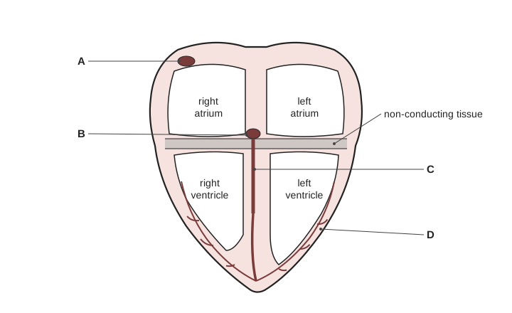
A vertical section through a heart seen from the front, with the right atrium and right ventricle on the left of the drawing and the left atrium and left ventricle on the right; the chambers are named. A grey band labelled non-conducting tissue separates the atria from the ventricles. Label A points to a small dark oval in the upper wall of the right atrium. Label B points to a small dark oval at the bottom of the wall between the two atria, just above the grey band. Label C points to a thick dark strand running down the wall between the two ventricles. Label D points to fine dark branching strands in the outer wall of the left ventricle, which run up from the apex.

Model answer

Starts each wave: A (sinoatrial node, SAN)
Delays the wave: B (atrioventricular node, AVN)
Carries the wave through the ventricle walls from the apex upwards: D (Purkyne fibres)

Explanation

The SAN in the wall of the right atrium sets the rhythm; its wave of electrical activity spreads across the atria, which contract. The non-conducting tissue stops the wave reaching the ventricles directly, so it passes through the AVN, which delays it, then down the bundle of His (C) in the septum to the apex and up through the Purkyne fibres in the ventricle walls. Confusing the bundle of His with the Purkyne fibres in the walls is a common error.

Mark scheme
  1. 1 mark

    A

    1 mark for each correct letter, in the order asked. If more than one letter is given for a structure, do not award that mark. If the answer names the structures instead of giving letters, credit a name only if it clearly matches the correct letter (sinoatrial node / SAN for A).

  2. 1 mark

    B

    B is the atrioventricular node (AVN). Accept AVN.

  3. 1 mark

    D

    D is the Purkyne fibres / Purkyne tissue in the ventricle walls. Accept Purkinje. Do not accept C (the bundle of His, which carries the wave down the septum to the apex).
Question 2

4 marks

In the heart, a layer of non-conducting tissue separates the atria from the ventricles. The wave of electrical activity from the atria reaches the ventricles only through the atrioventricular node (AVN), which delays it. The wave then passes down the bundle of His to the apex of the heart, and spreads up through the walls of the ventricles in Purkyne tissue.

Explain the importance of the non-conducting tissue, the delay at the AVN and the spread of the wave from the apex upwards.

Model answer

The non-conducting tissue stops the wave of electrical activity spreading straight from the atria into the ventricles, so the ventricles do not contract at the same time as the atria. The delay at the AVN gives the atria time to finish contracting, so the blood is emptied into the ventricles and the ventricles are full before they contract. Because the wave travels down to the apex and then up the ventricle walls, the ventricles contract from the bottom upwards, squeezing blood up and out into the aorta and pulmonary artery, which leave at the top of the heart; the Purkyne tissue spreads the wave quickly, so all the ventricle muscle contracts together.

Explanation

Each feature has a purpose in coordinating the heartbeat: the non-conducting tissue funnels the wave through the AVN, the AVN delay separates atrial and ventricular contraction, and conduction to the apex makes the ventricles contract from the bottom upwards towards the arteries. Explanations must say why the feature matters for pumping blood, not just describe the route of the wave.

Mark scheme
  1. 1 mark

    (Non-conducting tissue) stops the wave passing directly from the atria to the ventricles, so the ventricles do not contract at the same time as the atria

    Max 4. Accept 'so the wave can only reach the ventricles through the AVN'.

  2. 1 mark

    (Delay) allows the atria to (finish) contracting before the ventricles contract

    Accept 'atrial systole is complete before ventricular systole'.

  3. 1 mark

    (So) blood is emptied from the atria into the ventricles / the ventricles fill (fully) before they contract

  4. 1 mark

    (Apex upwards) the ventricles contract from the bottom / apex upwards, pushing blood up into the arteries / aorta and pulmonary artery (which leave at the top)

    Accept 'squeezes blood upwards towards the exits'.

  5. 1 mark

    Purkyne tissue spreads the wave rapidly, so all the ventricle muscle contracts at the same time / together (giving a forceful contraction)

    Accept 'simultaneous contraction of both ventricles'.

Question 3

5 marks

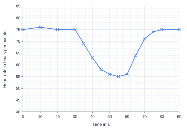
When the face is put into cold water, the heart rate decreases. This response is very strong in diving mammals such as seals. Scientists measured the heart rate of a volunteer who put her face into cold water from 30 s to 60 s. The graph shows the results.

Calculate the percentage decrease in heart rate from 30 s to the lowest heart rate recorded. Give your answer to 2 significant figures.

Cold receptors in the face send impulses to the medulla. Explain how this leads to the decrease in heart rate.

Suggest one advantage of this response to a diving seal.

Suggest one control the scientists should have used to show that the response was caused by the cold water.

Line graph of heart rate in beats per minute (y-axis, 40 to 85) against time in s (x-axis, 0 to 90). The line joins the points (0, 75), (10, 76), (20, 75), (30, 75), (35, 69), (40, 63), (45, 58), (50, 56), (55, 55), (60, 56), (65, 64), (70, 71), (75, 74), (80, 75), (90, 75).

Model answer

Heart rate at 30 s = 75 beats per minute; lowest = 55 beats per minute (at 55 s).
Percentage decrease = (75 − 55) ÷ 75 × 100 = 26.7 = 27%

The medulla (cardiovascular centre) sends more impulses along parasympathetic neurones in the vagus nerve to the SAN, which decreases the frequency of its waves of electrical activity, so the heart rate falls.

A slower heart rate means the heart uses less oxygen, so the seal's oxygen stores last longer while it is under water.

Control: repeat with the volunteer's face in water at body temperature (or holding her breath with her face out of the water), to show that the decrease is caused by the cold.

Explanation

Percentage decrease = decrease ÷ starting value × 100, so divide by 75, not by 55. The response is another reflex through the cardiovascular centre: more parasympathetic (vagus) impulses to the SAN slow the heart. A control must keep every other part of the procedure the same but remove the cold, so face immersion in warm water (or breath-holding alone) is suitable; simply repeating the experiment is not a control.

Mark scheme
  1. 1 mark

    Correct method: (75 − 55) ÷ 75 × 100; or a correct final answer of 27 (%) with no working

    Points 1 and 2 are the calculation: a correct answer of 27% with no working scores 2 marks. Accept for 1 mark: 26.7 (not given to 2 significant figures) OR 36 (difference divided by 55 instead of 75) OR 0.27 (not multiplied by 100).

  2. 1 mark

    27 (%)

  3. 1 mark

    More impulses (from the medulla / cardiovascular centre) along parasympathetic neurones / the vagus nerve to the SAN

    Accept 'fewer impulses along sympathetic neurones to the SAN'. The SAN must be named. Reject 'signals' or 'messages' for impulses, once only. Reject 'an impulse' or 'one impulse', once only (it is the frequency of impulses that changes). Accept action potentials for impulses.

  4. 1 mark

    Less oxygen is used (by the heart) / oxygen stores last longer while the seal is under water

    List rule: if more than one advantage is given, a wrong or contradictory extra answer cancels this mark. Accept 'so the seal can stay under water for longer'. Ignore 'saves energy' unqualified.

  5. 1 mark

    Repeat with the face in water at body temperature / warm water OR with the volunteer holding her breath without putting her face in water

    List rule: if more than one control is given, a wrong or contradictory extra answer cancels this mark. Ignore 'use more volunteers' (not a control).

Question 4

6 marks

Describe the roles of chemoreceptors and pressure receptors in controlling heart rate. Include their locations in your answer.

Model answer

Chemoreceptors are found in the aortic bodies and the carotid bodies. They detect a rise in the concentration of carbon dioxide (hydrogen ions) in the blood, which lowers its pH. Pressure receptors are found in the wall of the aorta and in the carotid sinus, and they detect changes in blood pressure.

Both types of receptor send impulses along sensory neurones to the cardiovascular centre in the medulla oblongata. When carbon dioxide rises (pH falls), or blood pressure falls, the centre sends more impulses along sympathetic neurones to the SAN, which increases the frequency of its waves of electrical activity, so the heart rate increases. When blood pressure rises, the centre sends more impulses along parasympathetic neurones in the vagus nerve to the SAN, which decreases the heart rate. The SAN, in the heart muscle, is the effector.

Explanation

The control pathway is receptor → sensory neurones → cardiovascular centre in the medulla → sympathetic (speeds up) or parasympathetic/vagus (slows down) neurones → SAN. Chemoreceptors respond to carbon dioxide and pH, not to oxygen, and a rise in carbon dioxide lowers pH. Write 'more impulses' or 'a higher frequency of impulses', not 'an impulse' or 'signals'.

Mark scheme
  1. 1 mark

    Chemoreceptors in the carotid bodies / carotid arteries OR in the aortic bodies / aorta

    Max 6. Either site is enough for this mark; both may be given. Reject 'signals' or 'messages' for impulses, once only. Reject 'an impulse' or 'one impulse', once only (it is the frequency of impulses that changes). Accept action potentials for impulses.

  2. 1 mark

    Chemoreceptors detect a rise in carbon dioxide / hydrogen ion concentration OR a fall in (blood) pH

    Reject 'detect a rise in pH'. Ignore 'detect oxygen'.

  3. 1 mark

    Pressure receptors in the aorta / aortic arch OR in the carotid sinus / carotid arteries

    Accept baroreceptors. Either site is enough for this mark; both may be given.

  4. 1 mark

    Pressure receptors detect changes in blood pressure

    Accept 'detect a rise or fall in blood pressure'.

  5. 1 mark

    (Receptors send) impulses (along sensory neurones) to the cardiovascular centre / cardiac centre in the medulla (oblongata)

    Accept 'to the medulla'.

  6. 1 mark

    A rise in carbon dioxide / fall in pH / fall in blood pressure leads to more impulses along sympathetic neurones to the SAN, so heart rate increases

    Accept 'fewer impulses along the vagus / parasympathetic neurones'.

  7. 1 mark

    A rise in blood pressure leads to more impulses along parasympathetic neurones / the vagus nerve to the SAN, so heart rate decreases

  8. 1 mark

    The SAN is the effector: it changes the frequency of waves of electrical activity (and so the heart rate)

    Accept 'cardiac muscle is the effector'.

Ready to try the rest? Every answer you write is marked against the mark scheme, so you can see exactly where the marks are.

Study Helper Hints first, answers after you try

Stuck on this one? Ask for a hint and I’ll point you in the right direction, without giving the answer away. Once you’ve checked your answer, I’ll show you how the marks are awarded.