
Level:
Correct: 0
Incorrect: 0
Marks: 0/0
Exam Practice
Course:
Topics:
AQA A-Level Biology (Paper 2) Smart practice A mixed round aimed at your weak spots
Unit 5
Energy transfers in and between organisms
Unit 6
Organisms respond to changes in their internal and external environments
Stimuli, both internal and external, are detected and lead to a response
Survival and response
0%
Receptors
0%
Control of heart rate
0%
Skeletal muscles are stimulated to contract by nerves and act as effectors
Skeletal muscles and muscle contraction
0%
Unit 7
Genetics, populations, evolution and ecosystems
Unit 8
The control of gene expression
Alteration of the sequence of bases in DNA can alter the structure of proteins
Gene mutations and their effects on proteins
0%
Using genome projects
0%
Differences in DNA between individuals of the same species can be exploited
DNA probes, screening and genetic counselling
0%
Genetic fingerprinting
0%
Unit 9
Comprehension
Avg
score:
–
Topics
done:
0/34

Gene expression and cancer

Practise 17 exam-style questions on Gene expression and cancer for AQA A-Level Biology (Paper 2). Write your answer as you would in the exam and get it marked in seconds against the mark scheme, point by point, with the Study Helper on hand if you get stuck.

  • 17 questions
  • 1–8 marks each
  • Instant marking
  • No sign-up needed
17 questions, marked as you go

Sample questions with model answers

Here are 4 of the questions in this topic, each with a model answer, an explanation of why it earns the marks and the mark scheme an examiner would use.

Question 1

3 marks

Doctors examined three tumours, J, K and L, removed from three different patients. The table shows some of their observations.

Which tumour is malignant? Give two pieces of evidence from the table that support your answer.

Tumour L was found inside the skull. Suggest why tumour L could be life-threatening even though it is not malignant.

Table with columns "Tumour", "Time for volume to double / days", "Capsule present", "Appearance of cells", "Cells in nearby lymph nodes". Tumour: J; Time for volume to double / days: 420; Capsule present: Yes; Appearance of cells: Like surrounding tissue; Cells in nearby lymph nodes: No | Tumour: K; Time for volume to double / days: 65; Capsule present: No; Appearance of cells: Unspecialised; irregular nuclei; Cells in nearby lymph nodes: Yes | Tumour: L; Time for volume to double / days: 350; Capsule present: Yes; Appearance of cells: Like surrounding tissue; Cells in nearby lymph nodes: No.

Model answer

Tumour K is malignant. It grows much faster than J and L: its volume doubles in 65 days, compared with 350 and 420 days. Also, cells from K have been found in nearby lymph nodes, so K has spread.

The skull cannot expand, so as tumour L grows it presses on the brain tissue (and its blood vessels), which can stop parts of the brain working.

Explanation

Malignant tumours grow rapidly, have no capsule, contain unspecialised cells with abnormal nuclei and can spread (metastasise) through the blood or lymph. Benign tumours grow slowly inside a capsule and do not spread, but they can still be dangerous when they press on vital organs such as the brain. Evidence must come from the table, so compare K with J and L rather than just listing features of malignant tumours.

Mark scheme
  1. 1 mark

    K grows faster / doubles in volume in a shorter time (65 days compared with 350 or 420 days)

    Max 1 if the answer does not identify K as the malignant tumour. Max 3. Points 1 to 4: 1 mark each, max 2 from these points. List rule: if more than two pieces of evidence are given, each wrong or contradictory extra piece cancels one correct piece. Accept 'K grows quickly' for point 1 only if it is linked to the doubling time in the table.

  2. 1 mark

    K has no capsule (so its cells are not contained / can invade nearby tissue)

    Accept 'K is not encapsulated'.

  3. 1 mark

    Cells of K are unspecialised / have large, irregular nuclei (unlike the surrounding tissue)

    Accept 'cells of K are not like the cells of the tissue around it'. Accept 'poorly differentiated'.

  4. 1 mark

    Cells from K are found in (nearby) lymph nodes, so K has spread / metastasised

    Accept 'K can spread to other parts of the body (through the lymph)'. Insufficient: 'K is in the lymph nodes' (the tumour itself is not in the lymph nodes).

  5. 1 mark

    L could press on / compress the brain (tissue) / blood vessels / nerves, because the skull cannot expand OR as it grows it could damage brain tissue and stop the brain working normally

    Accept 'there is no space for it to grow inside the skull, so it puts pressure on the brain'. Ignore 'it could become malignant' unless linked to pressure or damage. Reject 'L could spread to other parts of the brain' (L is benign).
Question 2

4 marks

Gene F codes for a receptor protein in the cell-surface membrane of skin cells. When a growth factor binds to this receptor, the receptor stimulates the cell to divide.

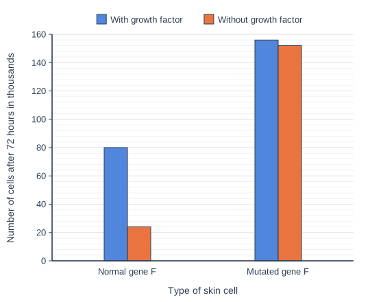
Scientists grew normal skin cells and skin cells with a mutated form of gene F, with or without the growth factor. Each culture started with 20 thousand cells. The chart shows the number of cells in each culture after 72 hours.

Calculate the mean rate of increase in the number of cells with the mutated gene F grown without the growth factor, in cells per hour. Give your answer to 2 significant figures.

The scientists concluded that the mutated form of gene F is an oncogene. Use the data to explain why.

Dual bar chart of number of cells after 72 hours in thousands for each type of skin cell, comparing With growth factor and Without growth factor (a key above the chart shows which bar is which). Normal gene F: With growth factor 80, Without growth factor 24; Mutated gene F: With growth factor 156, Without growth factor 152.

Model answer

Increase = 152 000 - 20 000 = 132 000 cells
Mean rate = 132 000 ÷ 72 = 1833 = 1800 cells per hour (2 s.f.)

Normal cells divided only when the growth factor was present: without it the number rose only from 20 to 24 thousand, compared with 80 thousand with it. Cells with mutated gene F reached about the same number with or without the growth factor (156 and 152 thousand). So the mutated receptor must be permanently active, stimulating division even when no growth factor is bound. Cell division is no longer controlled, which is what an oncogene does.

Explanation

Mean rate = change in number ÷ time: subtract the starting number (20 thousand) from the final number, divide by 72 hours, and remember that the chart is in thousands. Oncogenes are altered forms of genes that stimulate division; a mutation can produce a receptor that is active all the time, so the cell divides whether or not the signal (growth factor) is present. The evidence is the comparison: normal cells need the growth factor to divide, mutant cells do not.

Mark scheme
  1. 1 mark

    Correct method, e.g. (152 000 - 20 000) ÷ 72; or a correct final answer of 1800 / 1.8 × 10³ cells per hour with no working

    A correct answer of 1800 cells per hour (accept 1.8 × 10³ or 1.8 thousand cells per hour) with no working scores 2 marks (points 1 and 2). Accept for 1 mark: 1833 or 1833.3 (not given to 2 significant figures) OR 2100 or 2111 (152 000 ÷ 72, the starting number not subtracted) OR 1.8 with no indication that the unit is thousands of cells OR evidence of an increase of 132 000 (or 132 thousand) cells.

  2. 1 mark

    1800 / 1.8 × 10³ (cells per hour)

    Accept 1.8 thousand cells per hour. Do not accept 1.8 cells per hour.

  3. 1 mark

    Cells with mutated gene F divided (rapidly) without the growth factor / divided as much without it as with it (152 compared with 156 thousand), but normal cells hardly divided without it (20 to 24 thousand, compared with 80 thousand with it)

    The comparison must include both types of cell, with data or a clear comparative. Ignore 'grew' for divided if the numbers of cells are referred to.

  4. 1 mark

    The mutation (changes the tertiary structure so the) receptor is permanently active / stimulates division even when no growth factor is bound, so cell division is uncontrolled

    Accept 'the receptor is always switched on' only with reference to stimulating division without the growth factor. Reject 'no receptor is made' or 'the receptor is non-functional'.

Question 3

4 marks

Melanoma is a skin cancer. Allele R of a gene that affects skin pigment results in pale skin. Ultraviolet (UV) light is a mutagenic agent.

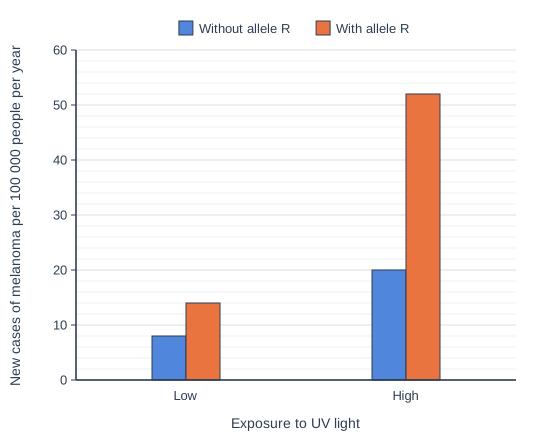
Scientists compared the incidence of melanoma in people with and without allele R who had low or high exposure to UV light. The chart shows their results.

What can you conclude about the effects of allele R and of UV exposure on the incidence of melanoma? Use the data to support your answer.

Suggest how this information could be used to reduce the incidence of melanoma.

Dual bar chart of new cases of melanoma per 100 000 people per year for each Exposure to UV light, comparing Without allele R and With allele R (a key above the chart shows which bar is which). Low: Without allele R 8, With allele R 14; High: Without allele R 20, With allele R 52.

Model answer

Allele R increases the incidence of melanoma: at low UV exposure, 14 per 100 000 compared with 8 without it. High UV exposure also increases it: from 8 to 20 per 100 000 in people without allele R. The effect of UV is much greater in people with allele R (14 rising to 52, an increase of 38, compared with an increase of 12), so the combined incidence (52) is much more than the separate effects would suggest (8 + 6 + 12 = 26): the genetic and environmental factors interact.

People with allele R, or pale skin, could be advised to avoid sunbeds and use sunscreen, and could be checked regularly for signs of melanoma.

Explanation

Here a genetic factor (allele R) and an environmental factor (UV, a mutagenic agent) both raise the risk, and together they raise it far more than either alone - people with pale skin absorb less UV in their pigment, so more UV reaches the DNA of their skin cells. Always support conclusions with figures. Understanding who is most at risk allows prevention to be targeted at them.

Mark scheme
  1. 1 mark

    Allele R increases the incidence of melanoma, e.g. from 8 to 14 per 100 000 at low UV exposure

    Max 4. Points 1 to 3: 1 mark each. Figures or a clear comparison are needed for each of points 1 to 3.

  2. 1 mark

    High UV exposure increases the incidence of melanoma, e.g. from 8 to 20 per 100 000 in people without allele R

  3. 1 mark

    The combined effect is greater than the sum of the separate effects / UV has a much bigger effect in people with allele R (an increase of 38 compared with 12 per 100 000; 52 is more than 8 + 6 + 12 = 26)

    Accept 'genetic and environmental factors interact' with supporting figures.

  4. 1 mark

    People with allele R / pale skin should be advised to avoid UV exposure / use sunscreen / avoid sunbeds OR be screened for allele R and checked regularly for skin changes

    Ignore 'avoid the sun' with no reference to people with allele R or pale skin; 'everyone should avoid UV' is insufficient.

Question 4

6 marks

Tumours develop when cell division is no longer controlled.

Explain how mutations and abnormal DNA methylation affecting oncogenes and tumour suppressor genes can result in the formation of a tumour.

Model answer

Tumour suppressor genes code for proteins that slow down cell division, or that cause cells with damaged DNA to die by apoptosis. A mutation in a tumour suppressor gene changes its base sequence, so the protein has a different amino acid sequence and tertiary structure and does not work. Increased methylation of the promoter of a tumour suppressor gene prevents its transcription, so the protein is not made at all.

Oncogenes code for proteins that stimulate cell division, such as receptors for growth factors. A mutation in an oncogene can make its protein permanently active, for example a receptor that stimulates division even when no growth factor is bound. Decreased methylation of an oncogene increases its transcription, so more of the protein is made.

In each case the brake on cell division is removed or the signal to divide is always on, so cells divide rapidly and uncontrollably by mitosis and form a tumour.

Explanation

Think of tumour suppressor genes as a brake and oncogenes as an accelerator: a tumour forms when the brake fails (inactivation by mutation or hypermethylation) or the accelerator is stuck on (activation by mutation or hypomethylation). Methylation is an epigenetic change: it alters transcription but not the base sequence, so it is not a mutation - a common error. Write about cell division, not 'growth', and about transcription, not genes being 'switched on'.

Mark scheme
  1. 1 mark

    Tumour suppressor genes (normally) code for proteins that slow down / inhibit cell division OR that cause cells with damaged DNA to die (apoptosis)

    Max 6. Ignore 'growth' for cell division throughout; ignore genes being 'switched on' or 'switched off' unless transcription or expression is also referred to correctly. Accept 'stop the cell cycle'.

  2. 1 mark

    A mutation in a tumour suppressor gene changes its base sequence, so the protein has a different amino acid sequence / tertiary structure and is non-functional / inactive

    Accept 'no functional tumour suppressor protein is produced'. Insufficient: 'the gene is mutated' with no effect on the protein.

  3. 1 mark

    Increased methylation / hypermethylation of a tumour suppressor gene (promoter) prevents its transcription, so the protein is not made

    Accept 'methyl groups added to the tumour suppressor gene stop RNA polymerase / transcription factors binding'. Do not award if methylation is described as a mutation or as changing the base sequence.

  4. 1 mark

    Oncogenes code for proteins that stimulate cell division

    Accept named examples, e.g. receptors for growth factors, or proteins that pass on a signal to divide. Accept proto-oncogene for oncogene throughout; the term is not required.

  5. 1 mark

    A mutation in an oncogene (proto-oncogene) makes its protein permanently active OR causes more of the protein to be produced / the gene to be permanently activated

    Accept 'the receptor is active even when no growth factor is bound'. Reject 'no protein is produced' or 'the protein is non-functional' for this point.

  6. 1 mark

    Decreased methylation / hypomethylation of an oncogene (promoter) increases its transcription / expression

    Do not award if methylation is described as a mutation or as changing the base sequence.

  7. 1 mark

    (The result is) rapid / uncontrolled cell division (by mitosis), forming a mass of cells / tumour

    Accept 'cell division cannot be regulated'. Reject meiosis.

Ready to try the rest? Every answer you write is marked against the mark scheme, so you can see exactly where the marks are.

Study Helper Hints first, answers after you try

Stuck on this one? Ask for a hint and I’ll point you in the right direction, without giving the answer away. Once you’ve checked your answer, I’ll show you how the marks are awarded.