Level:
Correct: 0
Incorrect: 0
Marks: 0/0
Exam Practice
Course:
Topics:
Avg
score:
–
Topics
done:
0/31

Properties of electromagnetic waves 2

Practise 17 exam-style questions on Properties of electromagnetic waves 2 for AQA GCSE Combined Science (Physics Paper 2). Write your answer as you would in the exam and get it marked in seconds against the mark scheme, point by point, with the Study Helper on hand if you get stuck.

  • 17 questions
  • 1–6 marks each
  • Instant marking
  • No sign-up needed
17 questions, marked as you go

Sample questions with model answers

Here are 4 of the questions in this topic, each with a model answer, an explanation of why it earns the marks and the mark scheme an examiner would use.

Question 1

2 marks

Describe how radio waves are produced by a radio transmitter.

Model answer

An alternating current in the transmitter circuit makes electrons oscillate in the aerial. These oscillations in the electrical circuit produce radio waves with the same frequency as the oscillations.

Explanation

Radio waves can be produced by oscillations in electrical circuits. Changing the frequency of the alternating current in the transmitter changes the frequency of the radio waves it sends out.

Mark scheme
  1. 1 mark

    by oscillations (of electrons / charges) in an electrical circuit

    allow an alternating current in the transmitter aerial

  2. 1 mark

    the radio waves have the same frequency as the oscillations (alternating current)

Question 2

2 marks

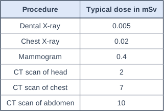
The table shows typical radiation doses from some medical procedures that use X-rays.

How many chest X-rays would give the same total dose as one CT scan of the abdomen?

Table with columns "Procedure", "Typical dose in mSv". Procedure: Dental X-ray; Typical dose in mSv: 0.005 | Procedure: Chest X-ray; Typical dose in mSv: 0.02 | Procedure: Mammogram; Typical dose in mSv: 0.4 | Procedure: CT scan of head; Typical dose in mSv: 2 | Procedure: CT scan of chest; Typical dose in mSv: 7 | Procedure: CT scan of abdomen; Typical dose in mSv: 10.

Model answer

Number of chest X-rays = 10 ÷ 0.02 = 500

Explanation

Divide the larger dose by the smaller one. A CT scan uses many X-ray images from different angles, so its dose, and therefore the risk of harm, is hundreds of times greater than a single chest X-ray.

Mark scheme
  1. 1 mark

    10 ÷ 0.02

    an answer of 500 scores 2 marks

  2. 1 mark

    500

Question 3

3 marks

The current in a radio transmitter's aerial oscillates with a frequency of 600 kHz. A radio receiver some distance away absorbs the radio waves.

Electromagnetic waves travel through air at 3.0 × 10⁸ m/s.

State the frequency of the alternating current produced in the receiver's aerial, and calculate the wavelength of the radio waves.

Model answer

The alternating current in the receiver has the same frequency as the radio waves, which is the same as the transmitter: 600 kHz.
f = 600 kHz = 600 000 Hz
v = f × λ
3.0 × 10⁸ = 600 000 × λ
λ = 3.0 × 10⁸ ÷ 600 000 = 500 m

Explanation

The oscillations in the transmitter produce radio waves of the same frequency, and absorbing those waves induces an alternating current of the same frequency again. Convert kHz to Hz before using the wave equation.

Mark scheme
  1. 1 mark

    frequency of the alternating current in the receiver = 600 kHz

    allow 600 000 Hz / 6.0 × 10⁵ Hz

  2. 1 mark

    3.0 × 10⁸ = 600 000 × λ

    allow λ = 3.0 × 10⁸ ÷ 6.0 × 10⁵

  3. 1 mark

    λ = 500 (m)

    an answer of 500 000 scores 1 mark for the calculation (kHz not converted)

Question 4

4 marks

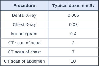
The table shows typical radiation doses from some medical procedures that use X-rays.

After a bad fall, a doctor advises a patient to have a CT scan of her head to check for serious injury. A dose of 2 mSv increases a person's lifetime risk of cancer by about 1 in 10 000.

The patient is worried about the risk. Use the information to evaluate whether she should have the scan.

Table with columns "Procedure", "Typical dose in mSv". Procedure: Dental X-ray; Typical dose in mSv: 0.005 | Procedure: Chest X-ray; Typical dose in mSv: 0.02 | Procedure: Mammogram; Typical dose in mSv: 0.4 | Procedure: CT scan of head; Typical dose in mSv: 2 | Procedure: CT scan of chest; Typical dose in mSv: 7 | Procedure: CT scan of abdomen; Typical dose in mSv: 10.

Model answer

A CT scan of the head gives a dose of 2 mSv, which is 100 times the dose from a chest X-ray. X-rays are ionising and can cause mutations in genes, so the scan does slightly increase her risk of cancer. However, the extra risk is only about 1 in 10 000 (0.01%), which is very small. A serious head injury could be life-threatening if it is not found, so the benefit of the scan is much greater than the risk. She should have the scan, but she should not have unnecessary repeat scans.

Explanation

Evaluating means weighing up both sides and reaching a judgement. Radiation dose measures the risk of harm, so compare the risk with the benefit of an accurate diagnosis.

Mark scheme
  1. 1 mark

    the CT scan of the head gives a much larger dose than a chest X-ray (100 times) or dental X-ray (400 times)

    max 4; full marks need a conclusion

  2. 1 mark

    X-rays are ionising and can cause mutation of genes, so there is some risk of cancer

  3. 1 mark

    but the extra risk is very small: 1 in 10 000 (0.01%)

    allow the dose is lower than the other CT scans in the table

  4. 1 mark

    a serious head injury could be life-threatening if it is not found and treated, so the benefit is much greater than the risk

  5. 1 mark

    conclusion: she should have the scan (but avoid unnecessary repeat scans)

    allow a reasoned conclusion either way if it is supported by the comparison of risk and benefit

Ready to try the rest? Every answer you write is marked against the mark scheme, so you can see exactly where the marks are.

Revision notes

Properties of electromagnetic waves 2

The big idea

Electromagnetic waves are generated and absorbed by changes in atoms and in the nuclei of atoms. At the high-frequency end of the spectrum, ultraviolet, X-rays and gamma rays can harm body tissue. How much harm depends on the type of radiation and the size of the dose, so you need to use dose data to judge risks.

Where electromagnetic waves come from

  • Changes in atoms: when an atom absorbs electromagnetic radiation, an electron moves to a higher energy level (further from the nucleus). When the electron falls back to a lower energy level, the atom emits electromagnetic radiation. These changes generate or absorb waves over a wide frequency range, including infrared, visible light, ultraviolet and X-rays.
  • Changes in the nucleus: gamma rays originate from changes in the nucleus of an atom. An unstable nucleus gives out its extra energy as a gamma ray.

X-rays and gamma rays can have the same wavelength, because their ranges of wavelength overlap. What tells them apart is where they come from: X-rays from changes involving electrons, gamma rays from the nucleus.

Higher tier only

Radio waves and electrical circuits. Radio waves can be produced by oscillations in electrical circuits. In a transmitter, an alternating current makes electrons oscillate up and down the aerial, and the aerial gives out radio waves with the same frequency as the oscillations.

When radio waves are absorbed by another aerial, they may create an alternating current with the same frequency as the radio wave itself. So radio waves can induce oscillations in an electrical circuit. This is how a radio or television receiver picks up a signal: a station broadcasting at 107 MHz produces a 107 MHz alternating current in the receiving aerial.

Hazards to the body

The electromagnetic spectrum drawn as a row of seven boxes, from left to right: box 1: radio waves; box 2: microwaves; box 3: infrared; box 4: visible light; box 5: ultraviolet (shaded); box 6: X-rays (shaded); box 7: gamma rays (shaded). Below the boxes, an arrow pointing right along the whole row is labelled "Increasing frequency". (Left to right the wavelength decreases and the frequency increases.)
The shaded groups can have hazardous effects on body tissue.
RadiationHazardous effects
UltravioletCan cause the skin to age prematurely and increase the risk of skin cancer (and sunburn)
X-raysIonising radiation: can cause the mutation of genes and cancer
Gamma raysIonising radiation: can cause the mutation of genes and cancer

Ionising radiation can knock electrons out of atoms, turning them into ions. In a cell this can damage DNA. X-rays and gamma rays also pass deep into the body, so they can reach internal organs, while ultraviolet is mostly absorbed by the skin. Lower-frequency waves, such as radio waves, are not ionising.

The effects depend on the type of radiation and the size of the dose.

Radiation dose

Radiation dose is a measure of the risk of harm resulting from an exposure of the body to the radiation. It is measured in sieverts (Sv), but you do not need to recall the unit.

\[1000 \text{ millisieverts (mSv)} = 1 \text{ sievert (Sv)}\]

To draw conclusions from dose data:

  • Convert to the same unit first: multiply by 1000 to go from Sv to mSv; divide by 1000 to go back.
  • Equal doses mean equal risk, whatever type of radiation they came from, because the dose already allows for the type.
  • Doses add up, so many small exposures can give a large total dose, and the risk grows with the dose.
  • Even a small dose carries a small risk. Doctors use X-rays only when the benefit of a diagnosis outweighs the risk.
  • The risk can be reduced: spend less time exposed, use shielding (lead absorbs X-rays and gamma rays), or use sunscreen and clothing against ultraviolet.

Reading a dosimeter

A technician who prepares gamma-emitting medical tracers wears a dosimeter badge. Over one year it records a dose of 0.0068 Sv. Her employer sets a limit of 20 mSv per year. What percentage of the limit did she receive, and what can you conclude?

Convert: 0.0068 Sv × 1000 = 6.8 mSv

Percentage of the limit: \(\dfrac{6.8}{20} \times 100 = 34\%\)

Her dose is about a third of the limit, so it is well within the limit her employer set. It still carries a small risk, which is why the dose is monitored and kept as low as possible.

Protection from ultraviolet

Arc welding gives out very intense ultraviolet. Explain why welders cover their arms and wear a mask.

  • Ultraviolet can make the skin age prematurely and increases the risk of skin cancer.
  • The harm depends on the dose. A welder is exposed for many hours, so without protection the dose would be large.
  • Clothing and the mask absorb the ultraviolet, so less reaches the skin and eyes. This lowers the dose and so lowers the risk.

Remember

  • Gamma rays come from changes in the nucleus; other electromagnetic waves come from changes in atoms.
  • Ultraviolet: premature ageing of the skin and a higher risk of skin cancer.
  • X-rays and gamma rays: ionising, causing gene mutation and cancer.
  • Radiation dose measures the risk of harm. 1000 mSv = 1 Sv.

Common mistakes

  • Saying radiation dose measures "how much radiation there is" or its energy. It measures the risk of harm to the body.
  • Converting the wrong way: 0.25 Sv is 250 mSv, not 0.000 25 mSv. The number in mSv is always the bigger one.
  • Mixing up the effects. Ultraviolet: premature ageing of the skin and skin cancer. X-rays and gamma rays: ionising, causing gene mutation and cancer.
  • Saying gamma rays come from electrons. They come from the nucleus.
  • Thinking all electromagnetic waves are equally harmful. Radio waves and visible light are far less hazardous than X-rays and gamma rays.

Exam tip

In "evaluate" questions about dose data, quote the numbers in the same unit, compare them (for example "40 times bigger"), say what that means for the risk, and finish with a judgement that weighs the risk against the benefit.

Study Helper Hints first, answers after you try

Stuck on this one? Ask for a hint and I’ll point you in the right direction, without giving the answer away. Once you’ve checked your answer, I’ll show you how the marks are awarded.