
Level:
Correct: 0
Incorrect: 0
Marks: 0/0
Exam Practice
Course:
Topics:
Edexcel GCSE Biology (Paper 2) Smart practice A mixed round aimed at your weak spots
Unit 1
Key concepts in biology
Unit 6
Plant structures and their functions
Unit 7
Animal coordination, control and homeostasis
Unit 8
Exchange and transport in animals
Unit 9
Ecosystems and material cycles
Avg
score:
–
Topics
done:
0/38

Core practical: microscopy and magnification

Practise 12 exam-style questions on Core practical: microscopy and magnification for Edexcel GCSE Biology (Paper 2). Write your answer as you would in the exam and get it marked in seconds against the mark scheme, point by point, with the Study Helper on hand if you get stuck.

  • 12 questions
  • 1–6 marks each
  • Instant marking
  • No sign-up needed
12 questions, marked as you go

Sample questions with model answers

Here are 4 of the questions in this topic, each with a model answer, an explanation of why it earns the marks and the mark scheme an examiner would use.

Question 1

1 mark

A student put a clear plastic ruler on the stage of a light microscope. She used it to find that the field of view was 2.0 mm wide at a total magnification of ×100.

She then looked at a thin slice of potato, using the same lenses. About 10 potato cells fitted in a line across the field of view.

What is the approximate length of one potato cell?

Choose your answer.

  • A 2 μm
  • B 20 μm
  • C 200 μm
  • D 20 mm

Model answer

C 200 μm

Explanation

The ruler gives the real width of the field of view, so dividing it by the number of cells that fit across it gives an estimate of the real length of one cell: 2.0 mm ÷ 10 = 0.2 mm = 200 μm. Dividing by the magnification as well is a common error. The answer is only an estimate, because the cells are not all the same size and do not fit exactly across the field of view.

Mark scheme
  1. 1 mark

    C / 200 μm (1)

    The only correct answer is C: the width of the field of view was measured with a ruler on the stage, so it is a real length; one cell = 2.0 mm ÷ 10 = 0.2 mm = 0.2 × 1000 = 200 μm. A is not correct because 0.2 mm has also been divided by the magnification (100), although the field of view is already a real length; B is not correct because 0.2 mm has been converted into micrometres using a factor of 100 instead of 1000; D is not correct because 2.0 mm has been multiplied by 10 instead of divided by 10. Accept the letter or the option. If more than one option is given, do not award the mark.
Question 2

2 marks

Chlorella is a single-celled alga that lives in ponds. A Chlorella cell has a diameter of about 6 μm.

A student wants to make a drawing of a Chlorella cell at a magnification of ×5000.

Calculate the diameter of the cell in her drawing, in mm.

Model answer

Size of image = size of real object × magnification
= 6 × 5000 = 30 000 μm
30 000 ÷ 1000 = 30 mm

Explanation

Rearranging magnification = size of image ÷ size of real object gives size of image = size of real object × magnification. Then convert micrometres into millimetres by dividing by 1000; dividing 6 by 5000 instead of multiplying gives an impossibly small drawing.

Mark scheme
  1. 1 mark

    (size of image = size of real object × magnification =) 6 × 5000 = 30 000 (μm) (1)

    An answer of 30 (mm) with no working scores 2 marks. accept 0.006 (mm) × 5000 (converting first) for this mark.

  2. 1 mark

    30 (mm) (1)

    accept 3 cm only if the unit is given. 30 000 (not converted into mm) scores 1 mark. award 1 mark for an answer with a power of ten error (e.g. 3 or 300) if the method (multiplying by 5000) is correct. 0.0012 (6 ÷ 5000) scores 0.

Question 3

3 marks

A student wants to look at the cells inside a leaf. She will cut very thin sections across the leaf using a sharp scalpel, and then look at them with a light microscope.

Describe how the student should cut the sections safely, and explain why the sections must be very thin.

Model answer

The student should cut the leaf on a white tile, holding it with forceps, and always cut away from her fingers so that the scalpel cannot cut her. The sections must be very thin so that light can pass through them, and so that there is only a single layer of cells, which do not overlap and can be seen clearly.

Explanation

A light microscope shines light through the specimen, so thick sections look dark and the cells overlap. Safety answers must say how the risk of cutting yourself is reduced, such as using a tile and cutting away from the fingers; 'be careful' alone does not gain credit.

Mark scheme
  1. 1 mark

    cut on a (white) tile / cutting board (1)

    Any three from the five points; max 3. Marking points 1 to 3 are for the safety precautions (max 2). ignore 'be careful' / 'use the scalpel carefully' alone.

  2. 1 mark

    cut away from the body / fingers (1)

    accept keep fingers away from the blade / behind the cutting edge.

  3. 1 mark

    hold the leaf with forceps / support the leaf in a holder (e.g. a piece of carrot or polystyrene) (1)

    accept do not hold the leaf directly in the fingers while cutting.

  4. 1 mark

    (thin) so that light can pass through the section (1)

    Marking points 4 and 5 are for the explanation (max 2).

  5. 1 mark

    so that the cells do not overlap / so that there is only one layer of cells, so individual cells can be seen (1)

    accept so the cells can be seen clearly / separately. ignore 'so it fits on the slide' / 'so it fits under the cover slip'.

Question 4

5 marks

A student cut a thin section across a leaf and looked at it with a light microscope. She made a drawing of one palisade cell and a drawing of one spongy mesophyll cell.

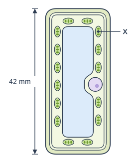
- Figure 1 shows her drawing of the palisade cell, with its length in the drawing marked. The magnification of this drawing is ×600. The structures labelled X were green.
- Her drawing of the spongy mesophyll cell is 48 mm long. The magnification of this drawing is ×1200.

The student wrote: 'The spongy mesophyll cell is longer than the palisade cell, because my drawing of it is longer.'

Name the structures labelled X.

Calculate the real length of each cell in micrometres (μm), and use your answers to evaluate the student's statement.

Diagram: a tall column-shaped cell with a thick outer layer and a thin line just inside it. A long pale blue space fills the centre. 15 small green oval structures with darker stacks inside are packed round the edge, and a round structure with a darker spot sits on the right-hand side. Label X points to one of the many small green oval structures with darker stacks inside, packed round the edge of the cell. A double-headed measuring arrow beside the cell spans its full height (the length of the whole cell), labelled "42 mm".

Model answer

X are chloroplasts.

Palisade cell: real length = size of image ÷ magnification = 42 ÷ 600 = 0.07 mm
0.07 × 1000 = 70 μm
Spongy mesophyll cell: real length = 48 ÷ 1200 = 0.04 mm
0.04 × 1000 = 40 μm

The student's statement is wrong. The palisade cell is really 70 μm long, which is 30 μm longer than the spongy mesophyll cell (40 μm). The drawing of the spongy mesophyll cell is longer only because it was drawn at twice the magnification, so the lengths of drawings made at different magnifications cannot be compared directly.

Explanation

Palisade cells are packed with chloroplasts, the green structures that contain chlorophyll. To compare real sizes, divide each image size by its own magnification and convert millimetres into micrometres by multiplying by 1000; the lengths of two drawings can only be compared directly if they have the same magnification. Multiplying by the magnification instead of dividing gives cells tens of metres long, a sign that the method is wrong.

Mark scheme
  1. 1 mark

    (X =) chloroplasts (1)

    Marking point 1 is for naming X. do not accept chlorophyll (the pigment, not the structure) / mitochondria / nuclei. Answers of 70 μm (palisade cell) and 40 μm (spongy mesophyll cell) with no working score marking points 2, 3 and 4. Marking point 5 is for the evaluation.

  2. 1 mark

    (real length of the palisade cell = 42 ÷ 600 =) 0.07 (mm) (1)

    real size = size of image ÷ magnification. accept 70 μm for this mark; accept 42 000 ÷ 600 (converting into μm first). 25 200 (42 × 600, multiplying by the magnification) does not gain this mark.

  3. 1 mark

    (real length of the spongy mesophyll cell = 48 ÷ 1200 =) 0.04 (mm) (1)

    accept 40 μm for this mark; accept 48 000 ÷ 1200 (converting into μm first). 57 600 (48 × 1200) does not gain this mark.

  4. 1 mark

    (conversion into μm, × 1000) 0.07 mm = 70 μm / 0.04 mm = 40 μm (1)

    this is the unit conversion mark: award it for a correct conversion of either length into μm; allow ecf from an incorrect length in mm. Lengths given only in mm (0.07 and 0.04) score marking points 2 and 3 only. If the lengths have a power of ten error from a correct method (e.g. 7 and 4, or 700 and 400 μm), award marking points 2 and 3 but not this mark.

  5. 1 mark

    the statement is wrong because the palisade cell is (really) longer than the spongy mesophyll cell (70 μm compared with 40 μm) (1)

    accept the palisade cell is 30 μm longer / 1.75 times as long; ecf: a conclusion that matches the student's own calculated lengths. The judgement must use the real lengths: 'the drawings were made at different magnifications, so they cannot be compared' alone does not gain this mark, but credit it as part of a correct comparison. ignore 'the student is wrong' with no comparison of the lengths.

Ready to try the rest? Every answer you write is marked against the mark scheme, so you can see exactly where the marks are.

Study Helper Hints first, answers after you try

Stuck on this one? Ask for a hint and I’ll point you in the right direction, without giving the answer away. Once you’ve checked your answer, I’ll show you how the marks are awarded.