
Level:
Correct: 0
Incorrect: 0
Marks: 0/0
Exam Practice
Course:
Topics:
OCR GCSE Physics (Paper 4) Smart practice A mixed round aimed at your weak spots
Unit 5
Waves in matter
Unit 6
Radioactivity
Unit 7
Energy
Unit 8
Global challenges
Unit 9
Practical skills
Avg
score:
–
Topics
done:
0/33

Waves for detecting hidden structures in the body

Practise 10 exam-style questions on Waves for detecting hidden structures in the body for OCR GCSE Physics (Paper 4). Write your answer as you would in the exam and get it marked in seconds against the mark scheme, point by point, with the Study Helper on hand if you get stuck.

  • 10 questions
  • 1–4 marks each
  • Instant marking
  • No sign-up needed
10 questions, marked as you go

Sample questions with model answers

Here are 4 of the questions in this topic, each with a model answer, an explanation of why it earns the marks and the mark scheme an examiner would use.

Question 1

1 mark

An ultrasound scanner makes images of the inside of the body from the echoes produced at boundaries between different materials. The table shows the percentage of the ultrasound energy that is reflected at three types of boundary.

Ultrasound scans are not used to make images of the inside of the lungs, which are full of air.

Which statement explains why?

Choose your answer.

  • A Air absorbs all of the ultrasound, so no echo at all is produced where the ultrasound reaches the lungs.
  • B Almost all of the ultrasound is reflected where it reaches the air, so very little passes into the lungs.
  • C Ultrasound cannot travel through a gas, because the particles in a gas are too far apart.
  • D Ultrasound travels faster in air than in soft tissue, so the echoes return too quickly.
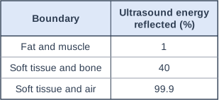
Table with columns "Boundary", "Ultrasound energy reflected (%)". Boundary: Fat and muscle; Ultrasound energy reflected (%): 1 | Boundary: Soft tissue and bone; Ultrasound energy reflected (%): 40 | Boundary: Soft tissue and air; Ultrasound energy reflected (%): 99.9.

Model answer

B

Explanation

The fraction of ultrasound reflected at a boundary depends on how different the two materials are. Between two soft tissues, such as fat and muscle, only about 1% is reflected, so the ultrasound can travel on to deeper boundaries; at a boundary with air, almost all of it is reflected. So the ultrasound cannot get into the lungs, and gas in the gut can also hide the organs behind it.

Mark scheme
  1. 1 mark

    B / almost all of the ultrasound is reflected where it reaches the air, so very little passes into the lungs

    The only correct answer is B: the table shows that 99.9% of the ultrasound energy is reflected at a boundary between soft tissue and air, so only about 0.1% passes into the lungs and there is almost nothing left to produce echoes from inside them. A is not correct because there is a very strong echo from the boundary with the air; C is not correct because sound can travel through gases, including air; D is not correct because ultrasound travels more slowly in air (about 330 m/s) than in soft tissue (about 1540 m/s). ALLOW the letter or the option. If more than one option is given, do not award the mark.

Question 2

2 marks

A doctor uses an infrared camera to examine a patient's painful knee. The image shows that the skin over one part of the knee joint is about 2 °C warmer than the skin around it.

Explain how the camera detects this warmer area, and suggest why the infrared image cannot show the shape of the bones inside the knee.

Model answer

All objects give out infrared, and warmer objects give out more infrared each second. The camera detects the extra infrared from the warmer skin over the joint, so that area shows up as a different colour on the image. The bones cannot be seen because infrared from inside the knee is absorbed by the tissues around it, so the camera only detects the infrared given out by the surface of the skin.

Explanation

An infrared camera does not send anything into the body: it detects the infrared that the skin itself emits, which depends on the temperature of the skin. Inflamed tissue is often warmer, so it shows up. Because body tissue absorbs infrared strongly, the image can only show surface temperatures, unlike X-rays or ultrasound, which pass into the body.

Mark scheme
  1. 1 mark

    warmer skin emits more infrared (radiation) each second / more intense infrared, which the camera detects, so this area shows up differently on the image

    ALLOW 'hotter objects give out more infrared'. DO NOT ALLOW 'the camera detects heat' with no reference to infrared radiation. DO NOT ALLOW 'the camera sends out infrared that is reflected by the warm area'.

  2. 1 mark

    infrared given out inside the knee is absorbed by the tissues around it / infrared does not pass through the skin and tissue, so the camera only detects infrared from the surface of the skin

    ALLOW 'infrared cannot travel far through the body'. IGNORE 'bones are cold'.

Question 3

3 marks

Doctors often check the brain of a newborn baby with an ultrasound scan. The probe is placed on the fontanelle, a soft area on the top of a baby's head where the bones of the skull have not yet joined. The fontanelle closes by the time a child is about 18 months old.

Ultrasound is not normally used to make images of the brain of an adult.

Explain why the scan is done through the fontanelle, and why this method cannot be used for an adult.

Model answer

When ultrasound meets bone, a large fraction of it is reflected at the boundary, and the bone also absorbs the ultrasound strongly, so very little gets through the skull. At the fontanelle there is no bone, so the ultrasound passes through soft tissue only and most of it reaches the brain. The echoes from boundaries inside the brain are then strong enough to make an image. In an adult the fontanelle has closed and the skull is bone all over, so too little ultrasound reaches the brain and returns to the probe.

Explanation

An ultrasound image needs ultrasound to reach the structure and enough of it to come back. Bone both reflects and absorbs ultrasound, so it acts like a barrier; the fontanelle is an 'acoustic window' through it. Strong answers explain what happens at the bone (reflection and absorption) and then link this to why the window is needed.

Mark scheme
  1. 1 mark

    at a boundary between soft tissue and bone, a large fraction of the ultrasound is reflected

    1 mark for each of the four points, max 3. ALLOW about 40% is reflected at a boundary between soft tissue and bone. ALLOW 'bone reflects most of the ultrasound'.

  2. 1 mark

    bone absorbs ultrasound strongly, so very little is transmitted through the skull

    ALLOW 'ultrasound cannot pass through the skull well'.

  3. 1 mark

    through the fontanelle the ultrasound passes only through soft tissue and liquid, so most of it is transmitted into the brain and the echoes from boundaries inside the brain are strong enough to detect

    ALLOW 'there is no bone in the way at the fontanelle'.

  4. 1 mark

    in an adult the fontanelle has closed / the skull is bone all over, so too little ultrasound reaches the brain and the echoes from the brain are too weak to form an image

    ALLOW the adult skull is thicker. DO NOT ALLOW 'adults are too big for ultrasound'.

Question 4

3 marks

Gallstones are small, hard stones that can form in the gallbladder, which is filled with a liquid called bile. Most gallstones are made mainly of cholesterol and contain very little calcium.

• Ultrasound travels at about 1500 m/s in bile, about 1540 m/s in the soft tissue of the gallbladder wall, and more than 2000 m/s in most gallstones.
• Most gallstones absorb X-rays by about the same amount as the bile and soft tissue around them.

On an ultrasound image, a gallstone shows up as a patch that is much brighter than the wall of the gallbladder.

Explain why most gallstones can be detected with ultrasound but not with an ordinary X-ray image, and why a gallstone appears much brighter than the gallbladder wall on the ultrasound image.

Model answer

An X-ray image shows differences in absorption. Most gallstones absorb X-rays by about the same amount as the bile and tissue around them, so about the same amount of X-rays reaches the detector behind a stone as behind the bile, and the stone does not show up.
An ultrasound image is made from echoes, and the fraction of ultrasound reflected at a boundary is larger when the speed changes more. Ultrasound travels at about 1500 m/s in bile but at more than 2000 m/s in a stone, so a large fraction is reflected at the surface of the stone, giving a strong echo and a bright patch. The speeds in bile and in the gallbladder wall (1500 m/s and 1540 m/s) are almost the same, so only a small fraction of the ultrasound is reflected there and most is transmitted, giving a weak echo, so the wall looks much dimmer.

Explanation

X-ray images rely on differences in absorption, while ultrasound images rely on reflection at boundaries, which is strongest where the speed (and density) changes most. A gallstone gives almost no X-ray contrast but a very strong ultrasound echo, which is why ultrasound is the usual test for gallstones. Strong answers compare the two boundaries using the speeds given.

Mark scheme
  1. 1 mark

    X-rays: the stone absorbs about the same amount of X-rays as the bile and tissue around it, so about the same amount of X-rays reaches the detector behind it / there is no contrast, so the stone does not show up

    DO NOT ALLOW 'X-rays pass straight through gallstones' with no comparison with the surrounding bile or tissue.

  2. 1 mark

    the speed of ultrasound changes a lot at the boundary between the bile and the stone (from about 1500 m/s to more than 2000 m/s), so a large fraction of the ultrasound is reflected there, giving a strong echo (a bright patch)

    ALLOW 'the stone is a solid in a liquid, so a lot of the ultrasound is reflected at its surface'. ALLOW a large difference in density. The idea of more reflection (a stronger echo) is needed.

  3. 1 mark

    the speeds in the bile and in the gallbladder wall are similar (1500 m/s and 1540 m/s), so only a small fraction of the ultrasound is reflected at that boundary, giving a weak echo (a dimmer image)

    ALLOW 'most of the ultrasound is transmitted from the bile into the wall'. ALLOW ORA (allow the reverse argument) when comparing the two boundaries.

Ready to try the rest? Every answer you write is marked against the mark scheme, so you can see exactly where the marks are.

Study Helper Hints first, answers after you try

Stuck on this one? Ask for a hint and I’ll point you in the right direction, without giving the answer away. Once you’ve checked your answer, I’ll show you how the marks are awarded.