
Level:
Correct: 0
Incorrect: 0
Marks: 0/0
Exam Practice
Course:
Topics:
Avg
score:
–
Topics
done:
0/27

Core practical: microscopy and magnification

Practise 15 exam-style questions on Core practical: microscopy and magnification for Edexcel GCSE Combined Science (Foundation Biology Paper 1). Write your answer as you would in the exam and get it marked in seconds against the mark scheme, point by point, with the Study Helper on hand if you get stuck.

  • 15 questions
  • 1–6 marks each
  • Instant marking
  • No sign-up needed
15 questions, marked as you go

Sample questions with model answers

Here are 4 of the questions in this topic, each with a model answer, an explanation of why it earns the marks and the mark scheme an examiner would use.

Question 1

1 mark

Students are asked to make a labelled scientific drawing of the cells they see with a light microscope.

Which of these describes a good scientific drawing?

Choose your answer.

  • A the darker parts of each cell are shaded in
  • B each part is coloured in a different colour
  • C the outlines are clear, unbroken lines made with a sharp pencil
  • D each label line ends in an arrowhead

Model answer

C the outlines are clear, unbroken lines made with a sharp pencil

Explanation

A scientific drawing records only what is seen, as clear pencil outlines with no shading or colour. Label lines are drawn with a ruler, do not cross and touch the part being labelled, with no arrowheads. The drawing should also have a title and the magnification.

Mark scheme
  1. 1 mark

    C / the outlines are clear, unbroken lines made with a sharp pencil

    The only correct answer is C: scientific drawings are made with a sharp pencil, using clear, continuous lines to show the outlines of the cells and their parts. A is not correct because scientific drawings are not shaded; B is not correct because scientific drawings are done in pencil, not in colour; D is not correct because label lines are straight lines drawn with a ruler, touching the part, with no arrowheads. Accept the letter or the option. If more than one option is given, do not award the mark.
Question 2

2 marks

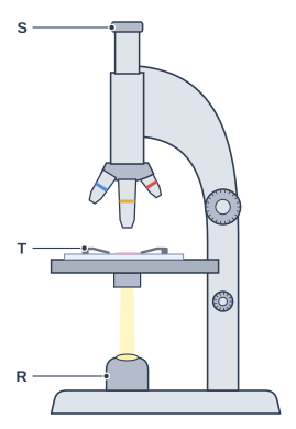
Three parts of the light microscope in the diagram are labelled R, S and T.

Give the letter of the part that:
- the student looks through
- holds the slide still on the stage
- shines light up through the slide.

Diagram of a light microscope, seen from the side. A light microscope seen from the side. A short, narrow tube sits on top of a wider vertical tube; below it a rotating disc carries three short tubes of different lengths pointing down. A thin strip of glass lies on a flat platform, held down by two small metal strips, above a small rounded housing with a bright window on the heavy flat bottom block. A large round knob and a smaller round knob are on the curved upright that joins the bottom block to the tubes. Label R points to the small rounded housing on the base, under the platform, with a bright window on top; label S points to the short, narrow tube at the very top; label T points to the small metal strips holding the glass strip down.

Model answer

The student looks through S (the eyepiece lens).
T (the clips) holds the slide still on the stage.
R (the lamp) shines light up through the slide.

Explanation

The eyepiece lens at the top is the lens you look through; the objective lenses on the turret are just above the slide. The clips hold the slide still on the stage, and the lamp in the base shines light up through the thin specimen so that it can be seen.

Mark scheme
  1. 2 marks

    looks through: S; holds the slide: T; shines light: R

    2 marks for all three letters correct; 1 mark for two letters correct. Accept the letters written in order (S, T, R) without the descriptions. Accept the letter with the name of the part, e.g. S (eyepiece lens), T (clips), R (lamp / light source); if a name contradicts its letter, credit the letter.

Question 3

2 marks

Nia looked at a slide of onion cells with a light microscope. With the ×10 objective lens she could see about 40 cells. When she changed to the ×40 objective lens, she could see only 2 or 3 cells, but each cell looked much bigger.

Explain why she could see fewer cells with the ×40 objective lens.

Model answer

The ×40 objective lens has a higher magnification, so each cell looks four times wider. This means that only a smaller area of the slide fits into the field of view, so she sees fewer cells.

Explanation

Magnification and field of view are linked: the higher the magnification, the smaller the area of the specimen that you can see at once. This is why cells are found with a low-power lens before changing to a higher power. Saying only that the cells look bigger does not explain why there are fewer of them.

Mark scheme
  1. 1 mark

    the ×40 lens gives a higher magnification / magnifies the cells (four times) more

    An explanation linking two points. accept each cell is made to look bigger / larger.

  2. 1 mark

    so a smaller area of the slide is seen / the field of view is smaller, so fewer cells fit into the view

    accept each cell takes up more of the field of view / you see less of the slide. Do not accept 'the cells are smaller' or 'the lens is closer to the slide' on its own.

Question 4

4 marks

Leo prepared a slide of onion skin cells. He put a piece of onion on a slide, added a drop of iodine solution and dropped a cover slip on top.

When he looked through the microscope, he saw:
- several large circles with thick black outlines
- areas where the cells looked dark because they were lying on top of each other.

Explain two ways Leo should change his method to get a clearer view of the cells.

Model answer

Leo should lower the cover slip slowly at an angle, using a mounted needle, so that no air bubbles are trapped. The black circles are air bubbles.
He should also peel off a thin layer of onion skin, just one cell thick, and lay it flat on the slide, so that light can pass through it and the cells do not overlap.

Explanation

Circles with thick black edges are air bubbles, which are avoided by lowering the cover slip from one side so that the liquid pushes the air out. Dark areas of overlapping cells mean the specimen was too thick or folded: a single, flat layer of cells lets light through so each cell can be seen. Because the command word is 'Explain', each change needs a reason to gain both of its marks.

Mark scheme
  1. 1 mark

    lower the cover slip slowly at an angle / lower it from one side using a mounted needle

    Two changes, each with a reason: max 4. Each reason mark depends on the change before it. accept use a mounted needle / forceps to lower the cover slip gently. Do not accept 'add the cover slip carefully' on its own.

  2. 1 mark

    so that no air bubbles are trapped (the black circles are air bubbles)

    accept to stop air bubbles forming / to push the air out.

  3. 1 mark

    use a thinner piece of onion / peel off a single layer of (epidermis) cells and lay it flat

    accept use the thin skin from the inside of the onion layer / spread it out with no folds / make sure the layer is one cell thick.

  4. 1 mark

    so that light can pass through it / so the cells do not overlap and each cell can be seen

    accept so that the cells are not on top of each other. Ignore 'so it is clearer' on its own.

Ready to try the rest? Every answer you write is marked against the mark scheme, so you can see exactly where the marks are.

Study Helper Hints first, answers after you try

Stuck on this one? Ask for a hint and I’ll point you in the right direction, without giving the answer away. Once you’ve checked your answer, I’ll show you how the marks are awarded.