
Level:
Correct: 0
Incorrect: 0
Marks: 0/0
Exam Practice
Course:
Topics:
OCR GCSE Biology (Paper 3) Smart practice A mixed round aimed at your weak spots
Avg
score:
–
Topics
done:
0/29

Light microscopes and staining

Practise 14 exam-style questions on Light microscopes and staining for OCR GCSE Biology (Paper 3). Write your answer as you would in the exam and get it marked in seconds against the mark scheme, point by point, with the Study Helper on hand if you get stuck.

  • 14 questions
  • 1–6 marks each
  • Instant marking
  • No sign-up needed
14 questions, marked as you go

Sample questions with model answers

Here are 4 of the questions in this topic, each with a model answer, an explanation of why it earns the marks and the mark scheme an examiner would use.

Question 1

1 mark

A student has a bottle of methylene blue stain. She wants to look at four different specimens with a light microscope.

For which specimen is a stain most needed to see the cells clearly?

Choose your answer.

  • A a leaf of Elodea pondweed
  • B a thin layer of skin from a red onion
  • C a thin slice of carrot
  • D cells scraped from the inside of the cheek

Model answer

D

Explanation

Stains are used to view colourless specimens. Cheek cells have no coloured structures, so a stain such as methylene blue is needed to give contrast; it colours the nucleus dark blue. The other three specimens already contain coloured structures or pigments.

Mark scheme
  1. 1 mark

    D / cells scraped from the inside of the cheek

    The only correct answer is D: cheek cells are almost colourless (transparent), so without a stain there is very little contrast and the cell outline and nucleus are hard to see. A is not correct because Elodea cells contain green chloroplasts, which can be seen without a stain; B is not correct because red onion skin cells have red-purple sap in their vacuoles, so the cells are already coloured; C is not correct because carrot cells contain orange pigment, so they are already coloured. ALLOW the letter or the specimen. If more than one option is given, do not award the mark.
Question 2

2 marks

A student has placed a thin layer of onion epidermis in a drop of water on a slide and lowered a cover slip over it.

She then puts a drop of iodine solution next to one edge of the cover slip, and touches a small piece of paper towel against the opposite edge of the cover slip.

Explain why she touches the paper towel against the opposite edge of the cover slip.

Model answer

The paper towel soaks up the water from under the cover slip at that edge. As the water is drawn out, the iodine solution is pulled in under the cover slip from the other edge and across the onion cells, so the cells are stained without lifting the cover slip.

Explanation

This is called irrigating the slide. The towel draws liquid out at one side, so the stain is pulled in from the other side and passes over the specimen. It lets a stain be added after the cover slip is in place, without trapping air bubbles. Answers that only say 'to dry the slide' do not explain how the stain reaches the cells.

Mark scheme
  1. 1 mark

    the paper towel absorbs / soaks up the water from under the cover slip

    max 2 from the three marking points. IGNORE just 'to dry the slide' or 'to clean the slide'. IGNORE references to removing air bubbles unless linked to staining without lifting the cover slip.

  2. 1 mark

    this draws / pulls the iodine solution under the cover slip / across the specimen

    ALLOW the iodine solution replaces the water under the cover slip. ALLOW so the stain spreads through / reaches all of the cells.

  3. 1 mark

    so the cells can be stained without lifting / removing the cover slip / without trapping air bubbles / without disturbing the specimen

    ALLOW AW, for example 'so the student does not have to make a new slide'.

Question 3

2 marks

A student looks at unstained cheek cells with a light microscope. With the lamp at full brightness the field of view is very bright and the cells are almost impossible to see.

The student turns down the brightness of the lamp. The outlines of the cells can now be seen.

Explain why turning down the brightness of the lamp makes the cells easier to see.

Model answer

Unstained cheek cells are almost colourless, so in very bright light they let nearly all the light through and look the same as the background. With less light there is more contrast between the cells and the background, so the cell outlines stand out.

Explanation

Transparent specimens show very little contrast. Reducing the light (or adding a stain) increases the contrast, so edges and structures become visible. It does not change the magnification or the resolution of the microscope, which is a common mistake.

Mark scheme
  1. 1 mark

    unstained cheek cells are (almost) colourless / transparent, so in bright light they look the same as the background

    ALLOW the cells let almost all the light through. ALLOW bright light 'washes out' the image of the cells.

  2. 1 mark

    with less light there is more contrast (between the cells and the background), so the cell outlines / edges stand out

    ALLOW AW for contrast, for example 'the cells look darker than the background'. ALLOW less glare / dazzle, so the faint cell outlines can be seen. IGNORE 'less glare' unqualified. DO NOT ALLOW 'less light increases the magnification' or 'increases the resolution'.

Question 4

4 marks

A student investigates how long onion epidermis needs to be left in iodine solution before the nuclei can be seen clearly.

She stains five pieces of epidermis for different times. For each piece she looks at 40 cells with a light microscope and counts how many of them have a nucleus that she can see clearly.

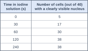
The table shows her results.

Describe and explain the pattern in the results.

Suggest the staining time the student should use in future, and justify your answer.

Table with columns "Time in iodine solution (s)", "Number of cells (out of 40) with a clearly visible nucleus". Time in iodine solution (s): 0; Number of cells (out of 40) with a clearly visible nucleus: 5 | Time in iodine solution (s): 30; Number of cells (out of 40) with a clearly visible nucleus: 17 | Time in iodine solution (s): 60; Number of cells (out of 40) with a clearly visible nucleus: 30 | Time in iodine solution (s): 120; Number of cells (out of 40) with a clearly visible nucleus: 38 | Time in iodine solution (s): 240; Number of cells (out of 40) with a clearly visible nucleus: 38.

Model answer

As the staining time increases from 0 s to 120 s, the number of cells with a clearly visible nucleus increases, from 5 to 38 out of 40. After 120 s there is no further increase: it is still 38 at 240 s. The longer the epidermis is left, the more iodine soaks into the cells and is taken up by the nuclei, so the nuclei become darker and stand out against the cytoplasm. By 120 s almost all the nuclei are stained. The student should use 120 s, because it is the shortest time that shows almost every nucleus; staining for longer does not make any more nuclei visible and wastes time.

Explanation

Describe the trend with data first (an increase, then a plateau), then explain it in terms of the stain giving the nucleus contrast. The best choice is the shortest time that gives the maximum result, here 120 s. A common mistake is to choose 240 s just because it is the longest time.

Mark scheme
  1. 1 mark

    the number of cells with a clearly visible nucleus increases as the staining time increases (from 0 s to 120 s)

    ALLOW correct use of data, for example 5 cells at 0 s rising to 38 cells at 120 s. ALLOW ORA (allow the reverse argument).

  2. 1 mark

    there is no further increase after 120 s / the number levels off at 38 (between 120 s and 240 s)

    ALLOW the number stays the same at 120 s and 240 s.

  3. 1 mark

    the longer the time, the more stain is taken up by the nuclei, so the nuclei are darker / contrast more with the cytoplasm

    ALLOW idea that the stain needs time to reach / soak into all of the cells. IGNORE 'the stain makes the nucleus bigger'. DO NOT ALLOW the stain magnifies the nucleus.

  4. 1 mark

    120 s (about 2 minutes), because it is the shortest time that makes (almost) all the nuclei visible / staining for longer gives no improvement

    The time and its justification are both needed for this mark. ALLOW a time between 120 s and 240 s if justified by the levelling off. DO NOT ALLOW 240 s justified only as 'the most cells'.

Ready to try the rest? Every answer you write is marked against the mark scheme, so you can see exactly where the marks are.

Study Helper Hints first, answers after you try

Stuck on this one? Ask for a hint and I’ll point you in the right direction, without giving the answer away. Once you’ve checked your answer, I’ll show you how the marks are awarded.