Level:
Correct: 0
Incorrect: 0
Marks: 0/0
Exam Practice
Course:
Topics:
Avg
score:
–
Topics
done:
0/23

The circulatory system, the heart and the blood

Practise 18 exam-style questions on The circulatory system, the heart and the blood for OCR GCSE Combined Science (Biology Paper 1). Write your answer as you would in the exam and get it marked in seconds against the mark scheme, point by point, with the Study Helper on hand if you get stuck.

  • 18 questions
  • 1–6 marks each
  • Instant marking
  • No sign-up needed
18 questions, marked as you go

Sample questions with model answers

Here are 4 of the questions in this topic, each with a model answer, an explanation of why it earns the marks and the mark scheme an examiner would use.

Question 1

1 mark

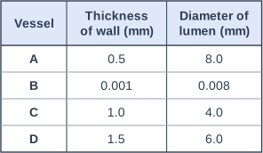
A scientist measured four blood vessels from the leg of a sheep. The table shows the results.

Which vessel is most likely to be a vein?

Choose your answer.

  • A vessel A
  • B vessel B
  • C vessel C
  • D vessel D
Table with columns "Vessel", "Thickness of wall (mm)", "Diameter of lumen (mm)". Vessel: A; Thickness of wall (mm): 0.5; Diameter of lumen (mm): 8.0 | Vessel: B; Thickness of wall (mm): 0.001; Diameter of lumen (mm): 0.008 | Vessel: C; Thickness of wall (mm): 1.0; Diameter of lumen (mm): 4.0 | Vessel: D; Thickness of wall (mm): 1.5; Diameter of lumen (mm): 6.0.

Model answer

A

Explanation

Compare the wall with the lumen: arteries have thick, muscular, elastic walls around a narrow lumen because they carry blood at high pressure; veins have thin walls and a wide lumen because the blood in them is at low pressure. A wall only one cell thick around a tiny lumen is a capillary.

Mark scheme
  1. 1 mark

    A / vessel A

    The only correct answer is A: vessel A has a thin wall (0.5 mm) and a wide lumen (8.0 mm), which is the structure of a vein carrying blood at low pressure. B is not correct because a wall 0.001 mm thick (one cell thick) and a lumen 0.008 mm wide are the features of a capillary. C is not correct because a thick wall (1.0 mm) around a narrow lumen (4.0 mm) is the structure of an artery. D is not correct because vessel D also has a thick wall (1.5 mm) compared with its lumen (6.0 mm), so it is a larger artery. ALLOW the letter or the option. If more than one option is given, do not award the mark.
Question 2

2 marks

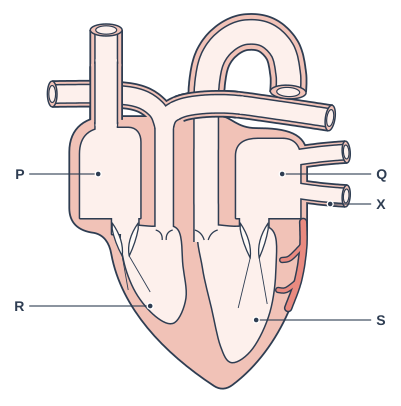
In the diagram of the heart, the four chambers are labelled P, Q, R and S. Blood returning from the lungs enters the heart through the vessels labelled X.

Give the letter of the chamber that this blood enters first.

Give the letter of the chamber that then pumps this blood out of the heart to the rest of the body.

Diagram of a vertical section through the heart, seen from the front. Four chambers are shown: two smaller upper chambers and two larger lower chambers, with a thick wall of muscle down the middle between the two lower chambers. The lower chamber on the right of the drawing has a much thicker muscular wall than the lower chamber on the left. Flaps held by thin threads lie between each upper chamber and the lower chamber below it, and small flaps sit at the bases of the two large vessels that leave the lower chambers. A large vessel enters the top of the upper left chamber; a vessel leaves the top of the lower left chamber and divides into two branches; two vessels enter the upper right chamber from the right; a large vessel leaves the top of the lower right chamber and arches over the top of the heart. A narrow branching vessel lies on the outer surface of the heart wall on the right. Label P points to the upper chamber on the left of the drawing; label Q points to the upper chamber on the right of the drawing; label R points to the lower chamber on the left of the drawing; label S points to the lower chamber on the right of the drawing, which has the thickest wall; label X points to one of the two vessels entering the upper right chamber from the right.

Model answer

The blood from the lungs (in the pulmonary veins, X) enters chamber Q, the left atrium, first.
Chamber S, the left ventricle, then pumps it out through the aorta to the rest of the body.

Explanation

Heart diagrams are drawn as if you are facing the person, so the left side of the heart is on the right of the diagram. Oxygenated blood from the lungs enters the left atrium, passes into the left ventricle and is pumped out through the aorta to the body. Chambers P and R, on the right side of the heart, deal with deoxygenated blood from the body.

Mark scheme
  1. 1 mark

    Q (the left atrium)

    ALLOW left atrium if no letter is given. DO NOT ALLOW P (the right atrium).

  2. 1 mark

    S (the left ventricle)

    ALLOW left ventricle if no letter is given. DO NOT ALLOW R (the right ventricle, which pumps blood to the lungs).

Question 3

2 marks

Carbon monoxide is a poisonous gas that can be released by faulty gas boilers. When it is breathed in, carbon monoxide combines with haemoglobin in red blood cells more strongly than oxygen does.

Explain why breathing in carbon monoxide can be dangerous.

Model answer

Carbon monoxide combines with the haemoglobin, so less haemoglobin is free to combine with oxygen and the red blood cells carry less oxygen. Less oxygen reaches the body cells, such as the brain cells, so they cannot carry out enough aerobic respiration to release the energy they need, and they can be damaged or die.

Explanation

Haemoglobin is what lets red blood cells carry oxygen. If carbon monoxide occupies it, the blood still flows normally but delivers much less oxygen, so cells cannot respire enough. A full answer links the haemoglobin to less oxygen transported and then to the effect on the cells.

Mark scheme
  1. 1 mark

    less haemoglobin is available to combine with / carry oxygen

    ALLOW the red blood cells carry less oxygen. ALLOW carbon monoxide takes the place of oxygen on the haemoglobin.

  2. 1 mark

    so less oxygen reaches the body cells / brain, so less (aerobic) respiration / cells cannot release enough energy (and may die)

    ALLOW the cells / tissues are starved of oxygen. IGNORE the person cannot breathe.

Question 4

3 marks

When a person starts to run, their heart beats faster and they breathe faster.

Explain how these two changes together help the leg muscles.

Model answer

During exercise the muscle cells respire faster, so they need more oxygen and glucose and they make more carbon dioxide. Breathing faster brings more oxygen into the alveoli, where it diffuses into the blood, and removes more carbon dioxide. The heart beating faster pumps the blood round faster, so more oxygen is carried from the lungs to the muscles every minute and more carbon dioxide is carried back to the lungs.

Explanation

The gas exchange system and the circulatory system work together: the lungs get oxygen into the blood and the heart moves the blood to where it is needed. Increasing only one of them would not be enough, because oxygen must both enter the blood faster and be delivered faster. Link each change to oxygen or carbon dioxide.

Mark scheme
  1. 1 mark

    the muscle cells respire faster, so they need more oxygen (and glucose) and make more carbon dioxide

    ALLOW the muscles need more energy (from respiration).

  2. 1 mark

    faster breathing brings more oxygen into the lungs / alveoli (for the blood to take in) and removes more carbon dioxide

    ALLOW more gas exchange in the lungs. IGNORE more air on its own.

  3. 1 mark

    a faster heart rate pumps blood around the body faster, so more oxygen is carried from the lungs to the muscles (and more carbon dioxide from the muscles to the lungs)

    ALLOW more blood reaches the muscles each minute. IGNORE the heart works harder on its own.

Ready to try the rest? Every answer you write is marked against the mark scheme, so you can see exactly where the marks are.

Study Helper Hints first, answers after you try

Stuck on this one? Ask for a hint and I’ll point you in the right direction, without giving the answer away. Once you’ve checked your answer, I’ll show you how the marks are awarded.