
Level:
Correct: 0
Incorrect: 0
Marks: 0/0
Exam Practice
Course:
Topics:
Avg
score:
–
Topics
done:
0/23

Light microscopes and staining

Practise 14 exam-style questions on Light microscopes and staining for OCR GCSE Combined Science (Biology Paper 1). Write your answer as you would in the exam and get it marked in seconds against the mark scheme, point by point, with the Study Helper on hand if you get stuck.

  • 14 questions
  • 1–6 marks each
  • Instant marking
  • No sign-up needed
14 questions, marked as you go

Sample questions with model answers

Here are 4 of the questions in this topic, each with a model answer, an explanation of why it earns the marks and the mark scheme an examiner would use.

Question 1

1 mark

Sudan III is a red stain. It colours fats and oils red, but it does not colour other substances.

A student puts a thin slice of a sunflower seed on a microscope slide and adds a drop of Sudan III.

Which structures in the seed cells will be coloured red?

Choose your answer.

  • A cell walls
  • B nuclei
  • C oil droplets
  • D starch grains

Model answer

C (oil droplets)

Explanation

Different stains colour different substances, so a stain can be chosen to highlight one type of structure. Sudan III colours fats and oils red, and the only oily structures in the seed cells are the oil droplets. Iodine solution would be used to show starch grains, and methylene blue to show nuclei.

Mark scheme
  1. 1 mark

    C / oil droplets

    The only correct answer is C: sunflower seed cells store oil as small droplets, and Sudan III colours only fats and oils, so only the oil droplets turn red. A is not correct because cell walls are made of cellulose, which is a carbohydrate, not a fat; B is not correct because nuclei are coloured by other stains, such as methylene blue, not by Sudan III; D is not correct because starch is a carbohydrate, which is shown by iodine solution (blue-black), not by Sudan III. ALLOW the letter or the option. If more than one option is given, do not award the mark.
Question 2

2 marks

A student puts a thin layer of cells from an unripe banana into a drop of water on a microscope slide. She lowers a cover slip on top of it.

She then puts a drop of iodine solution on top of the cover slip.

When she looks through the microscope, none of the starch grains in the banana cells have turned blue-black.

Explain why the cells were not stained, and describe how she should have added the iodine solution.

Model answer

The iodine solution stayed on top of the glass cover slip, so it could not reach the banana cells underneath. She should have added the drop of iodine solution to the banana cells before lowering the cover slip onto them.

Explanation

A cover slip is a thin piece of glass, and a stain cannot pass through glass. The stain must be in contact with the cells, so it is added to the specimen before the cover slip is lowered, or put at the edge of the cover slip so that it runs underneath.

Mark scheme
  1. 1 mark

    the iodine solution stayed on top of the cover slip / could not pass through the (glass) cover slip to reach the cells

    1 mark for point 1, and 1 mark for any one of points 2 and 3, max 2. If more than one method is given, a wrong one cancels a right one. ALLOW the cover slip was in the way / blocked the stain / kept the iodine away from the banana cells. IGNORE the stain did not work / there was not enough iodine.

  2. 1 mark

    add the iodine solution to the banana cells before lowering the cover slip

    ALLOW use iodine solution instead of the water / mix iodine solution with the water before adding the cover slip. ALLOW lift off the cover slip, add the iodine solution, then put the cover slip back. DO NOT ALLOW add more iodine solution on top of the cover slip / leave it for longer.

  3. 1 mark

    put a drop of iodine solution at the edge of the cover slip so that it runs under the cover slip (to the cells)

    ALLOW draw the iodine solution under the cover slip with a paper towel (held at the opposite edge).

Question 3

2 marks

Head lice lay eggs called nits on hair. A nit is about 0.8 mm long.

A student takes a photograph of a nit through a microscope. In the photograph the nit is 24 mm long.

Calculate the magnification of the photograph.

Model answer

magnification = image size ÷ real size
= 24 ÷ 0.8
= ×30

Explanation

Magnification = image size ÷ real size, with both sizes in the same unit (here both are in mm). 24 ÷ 0.8 = 30, so the photograph is 30 times bigger than the real nit. Dividing the real size by the image size gives a number less than 1, which cannot be right for a magnified image.

Mark scheme
  1. 1 mark

    (magnification = image size ÷ real size =) 24 ÷ 0.8; or a correct final answer of ×30 with no working

    An answer of ×30 (or 30) with no working scores 2 marks. DO NOT ALLOW 0.8 ÷ 24 (= 0.033) for any mark.

  2. 1 mark

    evaluation: (×) 30

    IGNORE units. ALLOW 30 times.

Question 4

4 marks

Hay fever forecasts are made by catching pollen grains from the air on sticky tape and identifying them with a light microscope.

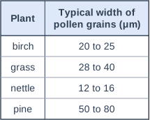
The table shows the approximate range of widths of the pollen grains of four plants.

A scientist photographs a pollen grain from the sticky tape. The magnification of the photograph is ×400. In the photograph the pollen grain is 13.1 mm wide.

Use the equation: real size = image size ÷ magnification

1 mm = 1000 μm

Calculate the real width of the pollen grain in μm. Give your answer to 2 significant figures.

Use your answer and the table to identify the plant that the pollen grain came from.

Table with columns "Plant", "Typical width of pollen grains (μm)". Plant: birch; Typical width of pollen grains (μm): 20 to 25 | Plant: grass; Typical width of pollen grains (μm): 28 to 40 | Plant: nettle; Typical width of pollen grains (μm): 12 to 16 | Plant: pine; Typical width of pollen grains (μm): 50 to 80.

Model answer

Real width = 13.1 ÷ 400 = 0.03275 mm
0.03275 × 1000 = 32.75 μm
Real width = 33 μm (to 2 significant figures)
The pollen grain came from a grass.

Explanation

Divide the image width by the magnification to get the real width in mm, then multiply by 1000 to change mm into μm: 13.1 ÷ 400 × 1000 = 32.75 μm, which is 33 μm to 2 significant figures. This is inside the range for grass pollen (28 to 40 μm) and no other plant's range. Check that you divided by the magnification: the real pollen grain must be much smaller than its image.

Mark scheme
  1. 1 mark

    13.1 ÷ 400 = 0.03275 (mm); or a correct final answer of 33 μm with no working

    An answer of 33 μm with no working scores marking points 1 to 3 only. ALLOW 32.75 μm or 32.8 μm for marking points 1 and 2 only (not rounded to 2 significant figures). ALLOW 0.033 mm for marking points 1 and 3 only (millimetres not converted to micrometres). ALLOW 13 100 ÷ 400 (image width changed into μm first). DO NOT ALLOW 13.1 × 400 for this mark or any later mark.

  2. 1 mark

    unit conversion: 0.03275 mm × 1000 = 32.75 (μm); or a correct final answer of 33 μm with no working

    ALLOW the conversion at any stage (for example 13.1 mm = 13 100 μm). ALLOW ECF from a value in mm that was found by dividing by 400.

  3. 1 mark

    evaluation: 33 (μm) (to 2 significant figures)

    ALLOW ECF for the rounding mark from a wrong answer correctly rounded to 2 significant figures (for example 0.033 mm when the conversion was missed). IGNORE units unless they contradict the answer.

  4. 1 mark

    grass

    ALLOW ECF: the plant in the table whose range of pollen widths includes (or is closest to) the width the student has calculated by dividing by 400 (in μm). DO NOT ALLOW a plant chosen with no calculated width, or from a width found by multiplying by 400.

Ready to try the rest? Every answer you write is marked against the mark scheme, so you can see exactly where the marks are.

Study Helper Hints first, answers after you try

Stuck on this one? Ask for a hint and I’ll point you in the right direction, without giving the answer away. Once you’ve checked your answer, I’ll show you how the marks are awarded.