
Level:
Correct: 0
Incorrect: 0
Marks: 0/0
Exam Practice
Course:
Topics:
OCR GCSE Biology (Paper 4) Smart practice A mixed round aimed at your weak spots
Avg
score:
–
Topics
done:
0/29

Defence against disease and the immune system

Practise 19 exam-style questions on Defence against disease and the immune system for OCR GCSE Biology (Paper 4). Write your answer as you would in the exam and get it marked in seconds against the mark scheme, point by point, with the Study Helper on hand if you get stuck.

  • 19 questions
  • 1–6 marks each
  • Instant marking
  • No sign-up needed
19 questions, marked as you go

Sample questions with model answers

Here are 4 of the questions in this topic, each with a model answer, an explanation of why it earns the marks and the mark scheme an examiner would use.

Question 1

1 mark

A patient has a very low number of lymphocytes in their blood. All the other components of their blood are present in normal numbers.

Which defence against pathogens is most likely to be reduced?

Choose your answer.

  • A clotting of the blood at a cut
  • B making antibodies against pathogens
  • C phagocytosis of bacteria
  • D trapping of pathogens in mucus

Model answer

B

Explanation

Each part of the blood has its own role in defence: phagocytes engulf and digest pathogens, lymphocytes make antibodies and antitoxins, and platelets help the blood to clot. Read the stem carefully: only the lymphocytes are reduced, so only the defences that depend on lymphocytes are affected.

Mark scheme
  1. 1 mark

    B / making antibodies against pathogens

    The only correct answer is B: lymphocytes are the white blood cells that make antibodies (and antitoxins). A is not correct because clotting depends on platelets, which are normal; C is not correct because phagocytosis is carried out by phagocytes, which are normal; D is not correct because mucus is made by cells lining the airways, not by blood cells. ALLOW the letter or the option. If more than one option is given, do not award the mark.
Question 2

2 marks

Stomach acid and antibodies are both part of the body's protection against pathogens.

Describe two differences between how stomach acid and how antibodies defend the body.

Model answer

Stomach acid is non-specific: it kills any type of pathogen in the food we swallow. Each antibody is specific: it binds only to one antigen, so it acts only on one type of pathogen.
Stomach acid works in the stomach, before pathogens enter the body tissues, but antibodies are made by lymphocytes and act on pathogens that have already got into the blood.

Explanation

Good answers make each difference a real comparison (acid ... but antibodies ...). The key ideas are specific versus non-specific, where each works, which cells make them and how they act. Avoid saying antibodies "fight" pathogens: say they bind to antigens or clump pathogens together.

Mark scheme
  1. 1 mark

    stomach acid is non-specific / acts on all types of pathogen, but each antibody is specific to one antigen / one type of pathogen

    1 mark for each difference, max 2. Each difference must refer to both stomach acid and antibodies, but ALLOW a comparison that is clearly implied (for example 'antibodies are specific to one antigen but stomach acid is not'). IGNORE 'antibodies fight pathogens'.

  2. 1 mark

    stomach acid kills pathogens in food and drink before they enter the body tissues / in the stomach, but antibodies act on pathogens that have entered the blood / the body tissues

  3. 1 mark

    stomach acid is made by cells in the stomach (lining), but antibodies are made by white blood cells / lymphocytes

  4. 1 mark

    stomach acid works in the same way every time, but antibodies are made faster (and in larger amounts) if the same pathogen enters the body again

    ALLOW antibody production involves memory cells but stomach acid does not.

  5. 1 mark

    stomach acid kills pathogens directly (by its low pH), but antibodies bind to antigens / clump pathogens together / help phagocytes to engulf the pathogens

    ALLOW acid denatures the proteins / enzymes of the pathogen.

Question 3

2 marks

Platelets stick to collagen fibres. In an undamaged blood vessel, the collagen fibres in the wall are covered by a smooth inner lining of cells. When a blood vessel is cut, the collagen fibres are exposed.

Suggest why it is important that platelets stick to collagen but do not stick to the lining of undamaged blood vessels.

Model answer

Platelets only stick where the collagen is exposed, so a clot forms only at a damaged vessel, where it is needed to stop bleeding and to seal the wound against pathogens. If platelets stuck to the lining of undamaged vessels, clots would form inside healthy vessels and block the blood flow, so the cells beyond the clot would be starved of oxygen and glucose (as in a heart attack or a stroke).

Explanation

Platelets are adapted to clot the blood only where it is needed. The trigger, exposed collagen, is present only at a wound. The best answers give both sides: clotting where it is needed, and the danger of clots blocking healthy vessels.

Mark scheme
  1. 1 mark

    so a clot forms only where a blood vessel is damaged / at a wound, (where it stops bleeding / stops pathogens entering)

    ALLOW platelets are not used up, so they are available to clot the blood at a wound.

  2. 1 mark

    (otherwise) clots would form inside undamaged blood vessels and block the flow of blood, so cells / tissues / organs would not get enough oxygen / glucose

    ALLOW a named effect of a blocked vessel, such as a heart attack or a stroke. IGNORE platelets would be wasted unless linked to fewer platelets being available at a wound.

Question 4

4 marks

A cannula is a thin plastic tube that is pushed through the skin into a vein, so that medicines can be given straight into the blood.

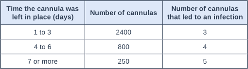
A hospital team recorded how long each cannula was left in place and whether it led to an infection. The table shows their results.

Calculate how many times greater the percentage of cannulas that led to an infection was when they were left in place for 7 days or more than when they were left in place for 1 to 3 days.

Use the data and your knowledge of the body's non-specific defences to explain why the risk of infection increases the longer a cannula is left in place.

Table with columns "Time the cannula was left in place (days)", "Number of cannulas", "Number of cannulas that led to an infection". Time the cannula was left in place (days): 1 to 3; Number of cannulas: 2400; Number of cannulas that led to an infection: 3 | Time the cannula was left in place (days): 4 to 6; Number of cannulas: 800; Number of cannulas that led to an infection: 4 | Time the cannula was left in place (days): 7 or more; Number of cannulas: 250; Number of cannulas that led to an infection: 5.

Model answer

1 to 3 days: 3 ÷ 2400 × 100 = 0.125 %
7 days or more: 5 ÷ 250 × 100 = 2.0 %
2.0 ÷ 0.125 = 16 times greater

Unbroken skin is a non-specific barrier that stops pathogens getting into the blood. A cannula passes through the skin, so bacteria from the surface of the skin can get into the blood along the tube. The longer the cannula is left in place, the longer the barrier is broken and the more time bacteria have to reproduce on the tube and get into the vein, so the percentage of cannulas that led to an infection rises from 0.125 % (1 to 3 days) to 0.5 % (4 to 6 days) and 2.0 % (7 days or more).

Explanation

Work out the percentage for each group before comparing them: the groups contain very different numbers of cannulas, so comparing the numbers of infections (5 and 3) gives the wrong answer. Then link the data to the skin as a non-specific barrier: a cannula breaks the barrier, and the longer it stays in, the more time bacteria have to get into the blood. This is why hospitals check cannulas every day and remove them as soon as they are no longer needed.

Mark scheme
  1. 1 mark

    working: 3 ÷ 2400 × 100 = 0.125 (%) and 5 ÷ 250 × 100 = 2.0 (%); or a correct final answer of 16 with no working

    An answer of 16 (times) with no working scores marking points 1 and 2. ALLOW the proportions 0.00125 and 0.02 for this mark. ALLOW 1 mark for 0.0625 (the percentages divided the wrong way round). DO NOT ALLOW 1.7 (5 ÷ 3, the numbers of infections compared instead of the percentages) for either mark.

  2. 1 mark

    evaluation: 16 (times)

    ALLOW 1.6 × 10¹.

  3. 1 mark

    unbroken skin is a barrier that stops pathogens getting into the blood / body, but the cannula passes through the skin, so pathogens (from the surface of the skin) can get into the blood (along the tube)

    ALLOW the cannula makes a hole / an opening in the skin barrier. IGNORE the cannula has germs on it unless linked to pathogens getting through the skin. DO NOT ALLOW the skin makes antibodies / white blood cells.

  4. 1 mark

    the longer the cannula is in place, the longer the skin barrier is broken / the more time pathogens have to get in / to reproduce on the tube, so the percentage of cannulas that led to an infection increases

    ALLOW bacteria grow along the (outside of the) tube into the vein over time. This mark needs the idea of more time for pathogens to get in or to reproduce. Data that may be quoted: 0.125 % (1 to 3 days), 0.5 % (4 to 6 days), 2.0 % (7 days or more); ALLOW ECF from the calculated percentages.

Ready to try the rest? Every answer you write is marked against the mark scheme, so you can see exactly where the marks are.

Study Helper Hints first, answers after you try

Stuck on this one? Ask for a hint and I’ll point you in the right direction, without giving the answer away. Once you’ve checked your answer, I’ll show you how the marks are awarded.