
Level:
Correct: 0
Incorrect: 0
Marks: 0/0
Exam Practice
Course:
Topics:
Avg
score:
–
Topics
done:
0/27

Microscopes, size and scale

Practise 14 exam-style questions on Microscopes, size and scale for Edexcel GCSE Combined Science (Biology Paper 1). Write your answer as you would in the exam and get it marked in seconds against the mark scheme, point by point, with the Study Helper on hand if you get stuck.

  • 14 questions
  • 1–6 marks each
  • Instant marking
  • No sign-up needed
14 questions, marked as you go

Sample questions with model answers

Here are 4 of the questions in this topic, each with a model answer, an explanation of why it earns the marks and the mark scheme an examiner would use.

Question 1

1 mark

Electron microscopes have a much higher resolution than light microscopes.

Resolution is the ability of a microscope to

Choose your answer.

  • A make the image larger
  • B show two close points as separate
  • C make the image brighter
  • D show a wider area of the specimen

Model answer

B show two close points as separate

Explanation

Resolution is how much detail can be seen: the higher the resolution, the closer together two points can be and still be seen as separate. Students often confuse resolution with magnification, but magnifying an image beyond the resolution limit only makes a blurred image bigger.

Mark scheme
  1. 1 mark

    B / show two close points as separate (1)

    The only correct answer is B: resolution is the smallest distance between two points that can still be seen as two separate points. A is not correct because this describes magnification; C is not correct because brightness depends on the light source, not the resolution; D is not correct because this describes the field of view. Accept the letter or the option. If more than one option is given, do not award the mark.
Question 2

2 marks

Light microscopes show mitochondria only as tiny dots or rods. Electron micrographs reveal that a mitochondrion has an inner membrane that is highly folded.

Suggest how this observation helped scientists to understand the role of mitochondria.

Model answer

The folds give the inner membrane a large surface area. This suggested that the membrane holds the enzymes for aerobic respiration, so the mitochondrion can carry out a lot of respiration and release energy quickly.

Explanation

Electron microscopes revealed internal detail that light microscopes cannot resolve, and the structure seen gave clues to the function: a large, folded membrane surface fits the role of mitochondria as the site of aerobic respiration. Answers must link the folds to surface area and to respiration, not just say that the folds 'help the mitochondrion work'.

Mark scheme
  1. 1 mark

    the folds give a large surface area (1)

    An explanation linking two from the three points; max 2. accept increases the surface area (of the membrane).

  2. 1 mark

    (for the) enzymes / reactions of (aerobic) respiration (1)

    accept for respiration to take place; must be linked to respiration, not photosynthesis.

  3. 1 mark

    so more respiration / more energy can be released (1)

    accept so the mitochondrion can release energy quickly; ignore makes energy.

Question 3

3 marks

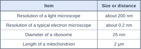
The table gives the resolution of two types of microscope and the sizes of two sub-cellular structures.

Mitochondria were first seen with light microscopes in the 1800s. Ribosomes were not seen until the 1950s, after the electron microscope had been developed.

Use the information in the table to explain why.

Table with columns "Item", "Size or distance". Item: Resolution of a light microscope; Size or distance: about 200 nm | Item: Resolution of a typical electron microscope; Size or distance: about 0.2 nm | Item: Diameter of a ribosome; Size or distance: 25 nm | Item: Length of a mitochondrion; Size or distance: 2 μm.

Model answer

A mitochondrion is 2 μm (2000 nm) long, which is ten times larger than the 200 nm resolution of a light microscope, so mitochondria could be seen with a light microscope. A ribosome is only 25 nm across, which is smaller than 200 nm, so a light microscope cannot resolve ribosomes as separate structures. The electron microscope can resolve details only 0.2 nm apart, so ribosomes could be seen once it was developed.

Explanation

Compare each size with the resolution, using the same units: 2 μm = 2000 nm. A structure smaller than the resolution limit cannot be distinguished, however much the image is magnified, which is why answers that only say 'electron microscopes magnify more' are not enough.

Mark scheme
  1. 1 mark

    a ribosome (25 nm) is smaller than the resolution of a light microscope (200 nm) (1)

    An explanation linking three from the four points; max 3. accept ribosomes are about 8 times smaller than the smallest detail a light microscope can show.

  2. 1 mark

    so ribosomes cannot be seen as separate structures / cannot be resolved with a light microscope (1)

    ignore 'ribosomes are too small to see' unless linked to resolution.

  3. 1 mark

    the electron microscope has a (much) higher resolution / can show details 0.2 nm apart, so ribosomes can be seen (1)

    accept the resolution of the electron microscope is 1000 times better; ignore greater magnification alone.

  4. 1 mark

    a mitochondrion (2 μm = 2000 nm) is larger than 200 nm, so it can be resolved with a light microscope (1)

    accept mitochondria are 10 times bigger than the resolution of a light microscope.

Question 4

4 marks

In 2016, scientists (Sender, Fuchs and Milo) estimated that the body of a typical 70 kg adult man contains about 3.0 × 10¹³ human cells and about 3.8 × 10¹³ bacterial cells.

Calculate the percentage of all the cells in the body that are bacterial cells.

The scientists also estimated that all of these bacterial cells together have a mass of only about 0.2 kg. Suggest why the bacterial cells have such a small total mass.

Model answer

Total number of cells = 3.0 × 10¹³ + 3.8 × 10¹³ = 6.8 × 10¹³
Percentage of bacterial cells = 3.8 × 10¹³ ÷ 6.8 × 10¹³ × 100 = 55.9%

Bacterial cells are prokaryotic cells, which are much smaller than human cells: a bacterium is about 1-2 μm long, whereas a human cell is usually about 10 μm or more across, so each bacterial cell has a tiny volume and mass compared with a human cell.

Explanation

To find a percentage of a total, first add the two numbers in standard form (the powers of ten are the same, so add 3.0 and 3.8), then divide the part by the total. A common error is to divide by the number of human cells, which gives 127%. More than half of the cells are bacteria, but they make up a tiny fraction of the mass because prokaryotic cells are about ten times smaller in length than most eukaryotic cells.

Mark scheme
  1. 1 mark

    (total number of cells =) 3.0 × 10¹³ + 3.8 × 10¹³ = 6.8 × 10¹³ (1)

    An answer in the range 55.8 to 56 % with no working scores marking points 1 and 2. Marking points 3 to 5 are for the suggestion: any two from the three; max 2. Total max 4.

  2. 1 mark

    (3.8 × 10¹³ ÷ 6.8 × 10¹³ × 100 =) 55.9 (%) (1)

    accept any value from 55.8 to 56 (e.g. 55.88 / 56); ecf from an incorrect total; 127 (%) (3.8 ÷ 3.0 × 100) scores 0; 44.1 (%) (the percentage of human cells) scores marking point 1 only.

  3. 1 mark

    (each) bacterial cell is much smaller / has a much smaller {volume / mass} than a human cell (1)

    accept prokaryotic cells are much smaller than eukaryotic cells; ignore 'bacteria are small / light' without a comparison with human cells.

  4. 1 mark

    a size comparison with values, e.g. a bacterium is about 1-2 μm long whereas most human cells are about 10 μm or more across / a bacterial cell has a volume hundreds of times smaller than a human cell (1)

    accept any sensible comparison of sizes or volumes (even a red blood cell, about 8 μm across, is much larger than a bacterium); accept bacteria are about ten times smaller in length.

  5. 1 mark

    bacteria are prokaryotic: they have no nucleus / no membrane-bound organelles, so each cell contains less material (1)

    accept human cells contain a nucleus, mitochondria and other organelles but bacterial cells do not; do not accept bacteria have no cell wall (they do have one).

Ready to try the rest? Every answer you write is marked against the mark scheme, so you can see exactly where the marks are.

Study Helper Hints first, answers after you try

Stuck on this one? Ask for a hint and I’ll point you in the right direction, without giving the answer away. Once you’ve checked your answer, I’ll show you how the marks are awarded.